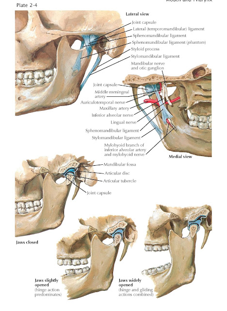
Temporomandibular Joint
The bony structures that enter into the formation of this joint are the head
of the mandibular condyle and the mandibular fossa and articular
tubercle of the temporal bone above. The head of the mandible is
ellipsoidal, with the long axis directed medially and slightly posteriorly.
This articular surface is markedly convex in the sagittal and coronal planes.
The articular surface on the temporal bone is concave posteriorly but becomes
more convex anteriorly. A fibrocartilage articular disk is inter- posed
between the two articular surfaces just described. Each surface of the disk
more or less conforms to the articular surface to which it is related, but the
shape of the disk between individuals is quite variable. The cartilage that
covers the bony articular surfaces differs from that of most joints in that it
is constituted from fibrocartilage tissue rather than hyaline cartilage,
although its gross appearance is similar to that of the articular cartilages of
other joints.
The temporomandibular joint is a true,
or synovial, joint, with two synovial spaces, one superior to the articular
disk and one inferior to it. This joint can be further described as having a hinge
motion in the lower space and a sliding motion in the upper space.
The capsular ligament is rather
loosely arranged, being attached superiorly to the margin of the articular
surface on the temporal bone and affixed inferiorly around the neck of the
mandible. The capsular ligament is firmly attached to the entire circumference
of the articular disk. Forming a pronounced thickening of the lateral aspect of
the capsule is the lateral temporomandibular ligament, which runs
inferiorly and posteriorly from the inferior border of the zygomatic process of
the temporal bone to the lateral and posterior sides of the neck of the
mandible. Two accessory ligaments are not blended with the capsule. The rather
thin sphenomandibular ligament runs from the spine of the sphenoid bone
to the lingula of the mandible, and the stylomandibular ligament, a
thickened band of deep cervical fascia, runs from the styloid process to the
lower part of the posterior border of the ramus of the mandible.
The temporomandibular joint receives
its nerve supply from the auriculotemporal and masseteric branches of
the mandibular division of the trigeminal nerve. Its arterial supply comes via
branches of the maxillary and superfi ial temporal arteries from the external
carotid artery.
The basic movements that are allowed
in the temporomandibular joint are (1) gliding of the articular disk anteriorly
and posteriorly on the articular surface of the temporal bone, accompanied by
the head of the mandible (which moves with the disk because the disk is
attached near the joint capsule’s attachment to the neck of the mandible and
the external pterygoid muscle is attached to both) and (2) the hinge movement
that takes place between the head of the mandible and the articu- lar disk. In
opening of the mouth, both movements are involved, with the hinge movement
predominating in slight opening and the gliding movement predominating in wide
opening. When chewing, one condyle remains more or less in position, while the
other moves backward and forward. This is combined with slight elevation and
depression of the mandible. If the mouth is opened just enough so that the
upper and lower incisor teeth can clear each other, the jaw can be protracted
and retracted, with the movement occurring in the upper joint.


