pediagenosis: Musculoskeletal
Article Update
Loading...
Showing posts with label Musculoskeletal. Show all posts

Sunday, May 18, 2025

INFECTIONS OF THE HAND

INFECTIONS OF THE HAND


INFECTIONS OF THE HAND

INFECTIONS OF THE HAND

Before the introduction of antibiotics, infections of the hand often led to prolonged morbidity, severe deformity, amputation, and even death. Kanavel’s classic article in 1939 on the pathways of purulent infection within the anatomic compartments of the hand opened the modern era of treatment for these problems. Although injuries in the industrial workplace are less prevalent than in Kanavel’s time, wounds of the hand still account for a large percentage of hand infections. A high incidence of hand infections is also associated with societal problems, such as intravenous injection of drugs with contaminated needles, wounds inflicted with various weapons in gang-related incidents, and complications of treatment with immunosuppressive agents. Human and animal bites may also have severe consequences.

Saturday, May 17, 2025

MUSCLES OF THE HAND

MUSCLES OF THE HAND


MUSCLES OF THE HAND

SPACES, BURSAE, AND TENDON SHEATHS OF THE HAND

INTRINSIC MUSCLES
The interosseous muscles occupy the intermetacarpal intervals and are of two types: dorsal and palmar. Each intermetacarpal space contains one palmar and one dorsal interosseous muscle. The four dorsal interosseous muscles are abductors of the digits and are bipennate; the three palmar interosseous muscles are adductors and are unipennate. The plane of reference for abduction and adduction of the fingers is the midplane of the third digit. This is evident on simultaneously spreading and then approximating the extended digits. The placement of these muscles follows from the above considerations of actions and reference plane for abduction and adduction.

Sunday, May 11, 2025

FLEXOR AND EXTENSOR TENDONS OF THE HAND

FLEXOR AND EXTENSOR TENDONS OF THE HAND


FLEXOR AND EXTENSOR TENDONS OF THE HAND

FLEXOR AND EXTENSOR TENDONS OF THE HAND

As the flexor and extensor tendons pass from the wrist to the hand, clinical zones have been described that help physicians articulate more precisely the significant anatomic differences that exist in each zone that affect finger function after injury (see Plates 4-5 and 4-6). As the extensor digitorum tendons diverge over the dorsum of the hand, they are interconnected by inter-tendinous connections. These prominently interconnect the tendons for the third, fourth, and fifth digits and severely limit the independent action of these digits, especially the fourth digit. Independent extensor action is retained for the index finger. The convergence of the tendon of the extensor pollicis longus muscle toward the tendons of the abductor pollicis longus and extensor pollicis brevis muscles defines a hollow known as the anatomic snuffbox (see Plate 4-14). In the floor of this hollow, the radial artery passes toward the dorsum of the hand and gives off its dorsal carpal branch.

Monday, April 28, 2025

Friday, April 25, 2025

REPLANTATION

REPLANTATION


REPLANTATION

REPLANTATION

Replantation is defined as the reattachment of a completely severed part. The first successful replantation of an above-elbow amputation was reported in 1962 by Malt and McLehman. In 1965, Komatsu and Tamai reported the successful replantation of a thumb. The development of this type of microsurgery has been greatly aided by advances in optical instrumentation and especially in the manufacture of needles and sutures fine enough to repair vessels 1 mm in diameter or less. Replantation is not suitable or possible for all patients with amputations. Great care must be given to the assessment of patients and their requirements. The surgical technique is exacting and the postoperative care prolonged and difficult. However, with an experienced team and a well-informed and motivated patient, the procedure can produce good functional and cosmetic results.

Sunday, April 20, 2025

DIABETIC FOOT ULCERATION

DIABETIC FOOT ULCERATION

DIABETIC FOOT ULCERATION

LESIONS OF THE DIABETIC FOOT

Patients with diabetes are susceptible to a host of foot-related problems. One of the most common and troublesome problems is ulceration and subsequent infection of the foot. Ulceration of the foot develops in the diabetic patient primarily as a result of peripheral neuropathy and loss of the normal protective sensation. Whereas the individual with normal protective sensation would immediately sense minor trauma such as the rubbing of a shoe and take immediate steps to correct it, the diabetic individual is not aware of the problem, allowing the pressure to continue unabated. Eventually, even minor repetitive trauma can result in formation of an ulcer. Ulcers occur most commonly on the weight-bearing plantar surface of the foot and over bony prominences. Once ulceration develops, it is also more likely to become infected in the diabetic patient owing to diminished immune function and impaired circulation. Failure to sense the normal signs of infection due to neuropathy can result in progression to osteomyelitis and extensive, limb-threatening infection in the diabetic patient.

SCOLIOSIS

SCOLIOSIS

SCOLIOSIS

PATHOLOGIC ANATOMY OF SCOLIOSIS

Scoliosis is a rotational deformity of the spine and ribs. While in most cases the cause of scoliosis is unknown (idiopathic scoliosis), in excess of 50 genetic markers have been identified as having a major role in adolescent idiopathic curves. Scoliosis may also result from a variety of congenital, neuromuscular, mesenchymal, and traumatic conditions, and it is commonly associated with neurofibromatosis.

Friday, November 22, 2024

KÖHLER DISEASE

KÖHLER DISEASE

KÖHLER DISEASE

KÖHLER DISEASE

Köhler disease is a self-limiting avascular necrosis of the tarsal navicular. It is usually unilateral and most often affects boys around age 4 and also girls around age 5. The navicular is located at the apex of the longitudinal arch of the foot, where it is subjected to repetitive compressive forces during weight bearing. Normally, the navicular is the last bone in the foot to ossify, and irregular ossification is not uncommon, especially in boys. The navicular ossifies later in boys than in girls, and delayed ossification appears to make the navicular more vulnerable to compressive damage.

Wednesday, November 20, 2024

COMMON FOOT INFECTIONS

COMMON FOOT INFECTIONS

COMMON FOOT INFECTIONS

COMMON INFECTIONS OF FOOT


The foot exists in an environment that unfortunately can be conducive to infection. Primarily, the use of shoes constricts the foot and produces a warm, moist environment that encourages bacterial growth. Foot infections can occur in all individuals. But, the diabetic patient is particularly susceptible to foot infection owing to the loss of protective sensation. Even trivial trauma either from a poorly fitting shoe or from bare-foot walking can result in violation of the skin and lead to severe infection. Poor blood supply and diminished immune function further compromise the diabetic patient’s ability to fight foot infection. Common locations for foot infections include the paronychial (nail) area and the deep spaces of the foot.

AMPUTATIONS IN THE FOOT

AMPUTATIONS IN THE FOOT

AMPUTATIONS IN THE FOOT

AMPUTATION OF FOOT
AMPUTATION OF FOOT


Amputation of all or a portion of the foot represents the most elemental form of foot surgery. Often it is disparaged by the surgeon, perhaps because it can be perceived as a failure of treatment. But when performed properly, amputation is truly a reconstructive procedure that can eradicate infection, correct deformity, decrease pain, and improve function.

Thursday, February 16, 2023

Motor Units, Recruitment and Summation

Motor Units, Recruitment and Summation


Motor Units, Recruitment and Summation.

Motor Units, Recruitment and Summation.


In normal skeletal muscle, fibres never contract as isolated individuals.
Several contract at almost the same time, as they are all supplied by the same α-motor neurone. The single motor neurone and all the fibres it innervates is called the motor unit (Fig. 14a). This is the smallest part of a muscle that can be made to contract independently of other parts of the muscle. The number of muscle fibres innervated by one motor unit can be as low as 5 or as high as 2000. The number is correlated with the precision with which the tension developed by the muscle is graded.
Atrophy

Atrophy


Atrophy


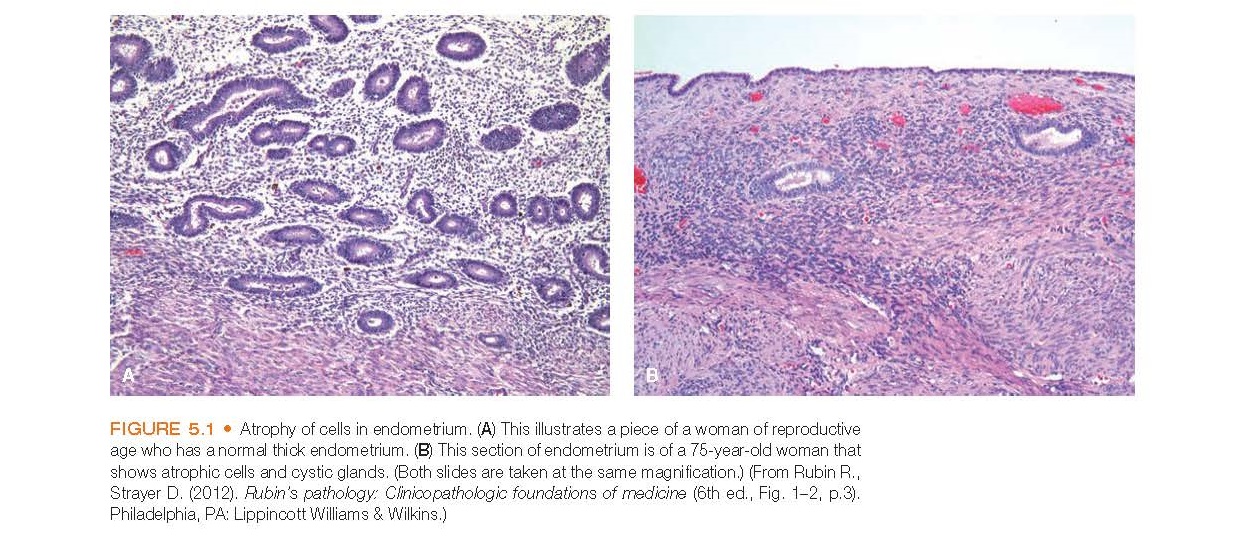
Atrophy of cells in endometrium. (A) This illustrates a piece of a woman of reproductive age who has a normal thick endometrium


When confronted with a decrease in work demands or adverse environmental conditions, most cells are able to revert to a smaller size and a lower and more efficient level of functioning that is compatible with survival. This decrease in cell size is called atrophy and is illustrated in Figure 5.1 regarding atrophy of the endometrium. Cells that are atrophied reduce their oxygen consumption and other cellular functions by decreasing the number and size of their organelles and other structures. There are fewer mitochondria, myofilaments, and endoplasmic reticulum structures. When a sufficient number of cells are involved, the entire tissue or muscle atrophies.

Monday, February 13, 2023

Connective or Supportive Tissue

Connective or Supportive Tissue


Connective or Supportive Tissue.


Diagrammatic representation of cells that may be seen in loose connective tissue. The cells lie in the extracellular matrix, which is bathed in tissue fluid that originates in the capillaries.

Connective or supportive tissue is the most abundant tissue in the body. As its name suggests, it connects and binds or supports the various tissues. Connective tissue is unique in that its cells produce the extracellular matrix that supports and holds tissues together. The capsules that surround organs of the body are composed of connective tissue. Bone, adipose tissue, and cartilage are specialized types of connective tissue that function to support the soft tissues of the body and store fat. The proximity of the extracellular matrix to blood vessels allows it to function as an exchange medium through which nutrients and metabolic wastes pass.

Wednesday, February 8, 2023

Muscle Tissue

Muscle Tissue


Muscle Tissue.


Connective tissue components of a skeletal muscle


Muscle tissue, whose primary function is contraction, is responsible for movement of the body and its parts and for changes in the size and shape of internal organs. Muscle tissue contains two types of fibers that are responsible for contraction: thin and thick filaments. The thin filaments are composed primarily of actin, whereas the thick filaments are composed of myosin. The two types of myofilaments occupy the bulk of the cytoplasm, which in muscle cells is called the sarcoplasm.

Sunday, February 5, 2023

Skeletal Muscle and its Contraction

Skeletal Muscle and its Contraction


Skeletal Muscle And Its Contraction

Skeletal Muscle And Its Contraction


Muscles make up about 50% of the adult body mass. There are three types of muscle: skeletal (muscle attached to the skeleton); cardiac (muscle involved in cardiac function; Chapter 15) (both of these are morphologically striated or striped and are commonly called striated muscles); and smooth (muscle involved in many involuntary processes in the blood vessels and gut; this type is not structurally striated, hence its name; Chapter 15). A comparison of the properties of the three muscle types is shown in Appendix I.

Friday, January 20, 2023

MYELODYSPLASIA

MYELODYSPLASIA

MYELODYSPLASIA

MYELODYSPLASIA

The number of infants with myelodysplasia who survive infancy has increased dramatically in the past 30 years, and as clinical experience with these patients has increased, new principles and techniques of treatment have emerged.

SPONDYLOLYSIS AND SPONDYLOLISTHESIS

SPONDYLOLYSIS AND SPONDYLOLISTHESIS

SPONDYLOLYSIS AND SPONDYLOLISTHESIS

SPONDYLOLYSIS AND SPONDYLOLISTHESIS

Spondylolysis may represent a stress fracture of the pars interarticularis of the fifth lumbar vertebra. When the fracture allows L5 to slip forward on S1, it is called isthmic spondylolisthesis. Dysplastic, or congenital, spondylolisthesis, in contrast, is due to anomalous development of the posterior structures of the lumbosacral junction.

LUMBOSACRAL AGENESIS

LUMBOSACRAL AGENESIS

LUMBOSACRAL AGENESIS

LUMBOSACRAL AGENESIS
Lumbosacral agenesis is a condition in which the sacrum and some of the lumbar vertebrae, or both, fail to develop. Although the etiology is not certain, it has been noted that 14% to 18% of patients have mothers with diabetes or a strong family history of diabetes.

Thursday, November 10, 2022

DISORDERS OF THE PATELLA

DISORDERS OF THE PATELLA
DISORDERS OF THE PATELLA

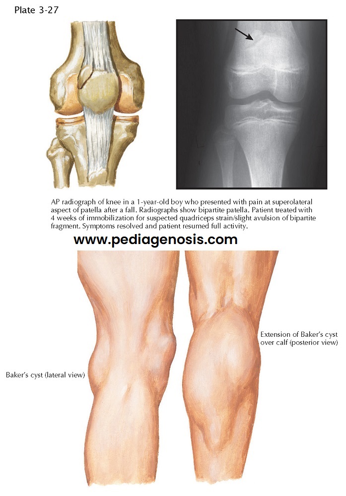
BIPARTITE PATELLA AND BAKER’S CYST


BIPARTITE PATELLA

Congenital fragmentation of the patella is relatively common. One type, bipartite patella, occurs in 1% to 2% of the population. This anatomic variant represents a true synchondrosis (a joint whose surfaces are connected by a cartilaginous plate). Most fragmented patellae remain asymptomatic, but, occasionally, direct trauma to the patella disrupts the synchondroses, causing symptoms that mimic those of a fracture.

Wednesday, September 21, 2022

REHABILITATION AFTER SPORTS INJURY

REHABILITATION AFTER SPORTS INJURY

REHABILITATION AFTER SPORTS INJURY

REHABILITATION AFTER SPORTS INJURY


The goal of conservative management of ligament injuries of the knee is to stabilize the action of the knee with the remaining uninjured, supportive structures. Rehabilitation must begin as soon as possible after injury, because disuse atrophy of the muscles occurs rapidly. Rehabilitation focuses on muscle strengthening, particularly strengthening of the extensor (quadriceps) muscles and the flexor (medial and lateral hamstring) muscles.

Anatomy Physiology

[AnatomyPhysiology][recentbylabel2]

Featured

[Featured][recentbylabel2]
Notification
This is just an example, you can fill it later with your own note.
Done