BLOOD SUPPLY OF THE UPPER
ARM
The subcutaneous veins of the limb are
interconnected with the deep veins of the limb via perforating veins.
Certain prominent veins, unaccompanied
by arteries, are found in the subcutaneous tissues of the limbs. The cephalic
and basilic veins, the principal superficial veins of the upper
limb, originate in venous radicals in the hand and digits.
Anastomosing longitudinal palmar
digital veins empty at the webs of the fingers into longitudinally oriented
dorsal digital veins. The dorsal veins of adjacent digits then unite to
form relatively short dorsal metacarpal veins, which end in the dorsal
venous arch. The radial continuation of the dorsal venous arch is the cephalic
vein, which receives the dorsal veins of the thumb and then ascends at the
radial border of the wrist. In the forearm, it tends to ascend at the anterior
border of the brachioradialis muscle, with tributaries from the dorsum of the
forearm. In the cubital space, the obliquely ascending median cubital vein connects
the cephalic and basilic veins (see Plate 2-12). Above the cubital fossa, the
cephalic vein runs in the lateral bicipital groove and then in the interval
between the deltoid and pectoralis major muscles, where it is accompanied by
the small deltoid branch of the thoracoacromial artery. At the deltopectoral
triangle, the cephalic vein perforates the costocoracoid membrane and empties
into the axillary vein. An accessory cephalic vein passes from
the dorsum of the forearm spirally laterally to join the cephalic vein at the
elbow.
The basilic vein continues to
the ulnar end of the venous arch of the dorsum of the hand. It ascends along
the ulnar border of the forearm and enters the cubital fossa anterior to the
medial epicondyle of the humerus. After receiving the median cubital vein, the
basilic vein continues upward in the medial bicipital groove, pierces the
brachial fascia a little below the middle of the arm, and enters the
neurovascular compartment of the medial intermuscular septum, where it lies
superficial to the brachial artery. In the distal axilla, it joins the brachial
veins to form the axillary vein.
The median antebrachial vein is
a frequent collecting vessel of the middle of the anterior surface of the
forearm (see Plate 2-12). It terminates in the cubital fossa in the median
cubital vein or in the basilic vein. It sometimes divides into a median
basilic vein and a median cephalic vein, which borders the biceps
brachii laterally and joins the cephalic vein. The median antebrachial vein may
be large or absent.
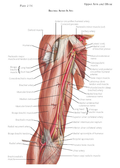
Brachial Artery
The brachial artery, the continuation
of the axillary artery, extends from the lower border of the teres major muscle
to its bifurcation opposite the neck of the radius in the lower part of the
cubital fossa. The course of the vessel may be marked with the limb in
right-angled abduction, when the vessel lies on a line connecting the middle of
the clavicle with the midpoint between the epicondyles of the humerus. The
brachial artery lies deep in the neurovascular compartment of the arm, flanked
by the brachial veins on either side and by the median nerve anterior to it.
The median nerve gradually crosses the artery to lie medial to it in the
cubital fossa. These structures are crossed by the bicipital aponeurosis at the
elbow.
The brachial artery is a single vessel
in 80% of cases. In the other 20% of cases, a superficial brachial artery
arises at the level of the upper arm and descends through the arm anterior to
the median nerve. Based on its forearm distribution, this artery is a high
radial artery in 10% of cases, is a high ulnar artery in 3%, and forms both
radial and ulnar arteries in 7%. In the last case, the brachial artery is
likely to become the common interosseous artery of the forearm.
The brachial artery provides numerous
muscular branches in the arm, principally from its lateral side. An especially
large branch supplies the biceps brachii muscle. The branches are named as
follows:
1. The deep
brachial artery arises from the medial and posterior aspect of the brachial
artery, below the tendon of the teres major muscle. It is the largest branch of
the brachial artery and accompanies the radial nerve in its diagonal course
around the humerus.
At the back of the humerus, the artery provides an ascending (deltoid)
branch, which reaches up to anastomose with the descending branch of the
posterior circumflex humeral artery. The deep brachial artery then divides into
the middle collateral artery and the radial collateral arteries. The middle
collateral artery plunges into the medial head of the triceps brachii
muscle and descends to the anastomosis of vessels at the level of the elbow.
The radial collateral artery continues with the radial nerve, both
perforating the lateral intermuscular septum to enter the anterior compartment. The artery ends in the elbow joint anastomosis, connecting in
particular with the radial recurrent artery from the radial artery. All these
branches nourish the muscles of the arm to which they are adjacent.
2. The nutrient
humeral artery arises about the middle of the arm and enters the nutrient canal
on the anteromedial surface of the humerus.
3. The superior
ulnar collateral artery arises from the brachial artery at or a little below
the middle of the arm. It pierces the medial intermuscular septum, descending behind
it with the ulnar nerve. With the nerve, it passes behind the medial epicondyle
of the humerus to anastomose with the inferior ulnar collateral artery and the posterior
ulnar recurrent branch of the ulnar artery.
4. The inferior
ulnar collateral artery arises from the brachial artery about 3 cm above the
medial epicondyle. It divides on the brachialis muscle into anterior and posterior
branches. Both these branches reach the anastomosis around the elbow joint, anterior
and posterior to it, respectively.
Brachial veins accompany the artery,
one on either side of it. They are formed from the venae comitantes of the
radial and ulnar arteries and have tributaries that accompany the branches of
the brachial artery, draining the areas supplied by the arteries. The brachial
veins contain valves and frequently anastomose with one another. At the lower
border of the teres major muscle, the lateral of the two veins crosses the artery
to join the more medial one; then, joined by the basilic vein, they form the
axillary vein.
Blood Supply To The Elbow
The blood supply of the elbow joint
comes from the anastomosis of the collateral branches of the brachial artery
and the recurrent branches of the radial and ulnar arteries.
Cubital Fossa
Like the axilla, the cubital fossa is
a space at the bend of the elbow where it is helpful to note the important
relationships of structures that overlie the elbow joint. It is described as a
triangular space, apex downward, and is bound above by a line connecting the
epicondyles of the humerus. The converging side borders are muscular, the
pronator teres muscle medially and the brachioradialis muscle laterally. The
floor of the space is also muscular, consisting of the brachialis muscle of the
arm and the supinator muscle of the forearm; deep to these muscles is the elbow
joint.
The readily palpable tendon of the
biceps brachii muscle descends centrally through the space, and its bicipital
aponeurosis spans medialward across the brachial artery and median nerve to
blend with the forearm fascia over the flexor muscle mass. Directly medial to
the biceps brachii tendon, the brachial artery divides into the radial
and ulnar arteries in the inferior part of the cubital fossa
opposite the neck of the radius.
Although submerged between the
brachioradialis and brachialis muscles, the radial nerve can be exposed
by drawing the brachioradialis muscle lateralward and can be followed to its
bifurcation into deep and superficial branches. Superficially, the medial
cubital vein crosses obliquely, overlying the bicipital aponeurosis; and a medial
cephalic vein may, on occasion, lie subcutaneously toward the lateral side
of the fossa. The medial antebrachial cutaneous nerve crosses the median
cubital vein, and the lateral antebrachial cutaneous nerve passes deep to
the median cephalic vein, if it is present.


