URETERAL DUPLICATION
As shown in Plate 2-1, the ureteric buds appear toward the caudal ends of
the mesonephric ducts at 5 weeks of gestation. Each bud grows into its adjacent
mass of metanephric mesenchyme, the precursor of the kidney, to form a ureter,
a pelvicalyceal system, and collecting ducts.
Ureteral duplication results from
abnormalities of the ureteric bud. It is one of the most common congenital
malformations of the urinary tract, with an incidence of approximately 1 in
125. Duplication is more often unilateral than bilateral, and more often
incomplete than complete. There does not appear to be any predilection for a
particular side.
Complete Ureteral Duplication
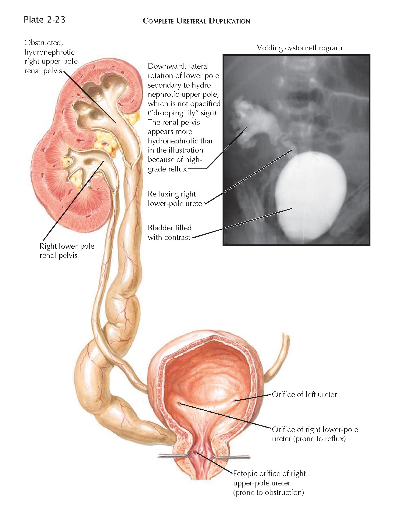
In complete ureteral duplication, the
kidney is drained by two distinct renal pelves, each of which leads to a ureter
with its own insertion into the bladder. This anomaly occurs when a single
mesonephric duct sprouts two ureteric buds, each of which induces a separate
portion of the adjacent metanephric mesenchyme. The more cranial of the two
ureteric buds becomes the collecting system of the upper pole, while the more
caudal of the ureteric buds becomes the collecting system of the lower pole.
Because of the manner in which the mesonephric ducts exstrophy into the
bladder; however, the upper pole ureter terminates at an orifice located
inferior and medial to that of the lower pole ureter. In many cases, the upper
pole ureter has an ectopic site of termination, reflecting an especially cranial
position of the ureteric bud from which it originated. The consistent pattern
of ureteral crossing seen in a duplicated system, where the ureter serving the
upper pole terminates inferior to the ureter serving the lower pole, is known
as the Weigert-Meyer law.
The upper-pole collecting system tends
to serve about one third of the renal parenchyma. The upper pole ureter
generally has a long course within the bladder wall, frequently has an ectopic
site of termination, and is also prone to ureterocele. Because of these
factors, upper pole obstruction and hydroureteronephrosis is common.
Meanwhile, the lower pole ureter tends
to have a short course within the bladder wall because of its superior and
lateral site of termination. As a result, it is prone to vesicoureteral reflux
(VUR, see Plate 2-21), which can cause lower pole hydronephrosis if severe.
Complete ureteral duplication may be
detected using various imaging techniques, including intravenous pyelography,
ultrasound, CT, and renal scanning. A classic finding is known as the “drooping
lily” sign. It consists of downward and lateral rotation of the lower pole
segment by an obstructed, hydronephrotic, poorly functioning (and thus
nonopacified) upper pole segment. If the lower pole reflux is marked enough, the
drooping lily sign may be seen on voiding cystourethrogram (VCUG).
Several different patterns of
incomplete ureteral duplication may be seen, reflecting various embryologic anomalies.
Unlike completely duplicated ureters, incompletely duplicated ureters rarely
cause obstruction, VUR, or other sequelae. As a result, they are generally
asymptomatic and discovered as incidental findings.
A bifid ureter, or
“Y ureter,” occurs when one ureteric bud arises from the mesonephric duct but
divides before entering the metanephric mesenchyme. As a result, the upper and
lower poles are drained by different renal pelves, but the associated ureters
fuse before the ureterovesical junction.
The site of ureteral fusion can occur
at any distance from the bladder. In some cases, the ureters do not fuse until
the ureterovesical junction, where they form a short common ureteral trunk.
A blind-ending ureter is a rarer kind of incomplete duplication. As with
a bifid ureter, this anomaly reflects early division of a single ureteric bud;
however, in this case, one of the ureteric bud branches fails to induce a
portion of metanephric mesenchyme. As a result, a bifid ureter is seen, but one
branch does not drain any renal parenchyma and is thus blind-ending. For
unknown reasons, blind-ending ureters occur three times more often in women and
are more often found on the right side.
The blind-ending ureteral branch
typically terminates adjacent to the distal or middle section of the normal
branch. It contains all of the normal layers of the ureteral wall, and it
terminates either as a dilated bulb or as an atretic stalk.
Because the blind-ending ureter does
not drain renal parenchyma, it cannot be seen on excretory studies, such as
intravenous pyelogram or contrast-enhanced CT. As a result, retrograde studies
are generally required to establish the diagnosis.
An “inverted Y” ureter is the rarest form of incomplete duplication. It
occurs when two ureteric buds appear on the mesonephric duct but fuse before
reaching the metanephric mesenchyme. As a result, a single renal pelvis is
seen, but the ureter divides as it approaches the bladder, such that two
distinct ureteric orifices are seen on the affected side. One of the ureteral
branches often has an ectopic orifice or forms a ureterocele, resulting in
obstruction.


