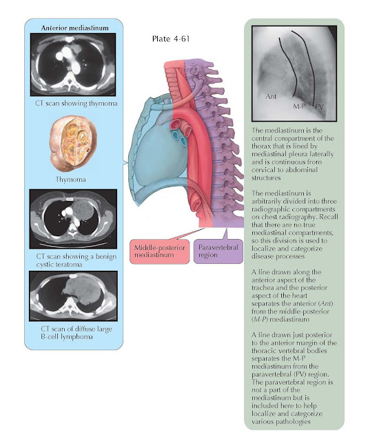
MEDIASTINAL TUMORS: ANTERIOR
MEDIASTINUM
The mediastinum is the central space in the chest cavity that is bounded
by the sternum anteriorly, the pleura of the lungs laterally, the vertebrae
posteriorly, the thoracic inlet superiorly, and the diaphragm inferiorly. The
mediastinum does not have rigid structures that divide it into compartments,
but for anatomic and clinical purposes, it is generally divided into anterior,
middle- posterior, and paravertebral compartments. It should be noted that the
paravertebral regions do not form part of the mediastinum proper. The anterior
mediastinum is defined by an imaginary line drawn along the anterior trachea and
posterior cardiac border on a lateral chest radiograph. The most common tumors
of the anterior mediastinum are thymoma, lymphoma, germ cell tumors, and
thyroid (goiter). Cystic hygroma should also be considered, usually in
children.
Tumors of the anterior mediastinum account for 50% of
all mediastinal tumors. They may be asymptomatic at diagnosis, or patients may
complain of cough, dyspnea, or vague chest discomfort. Thymoma is the most
common mediastinal tumor in adults. From 30% to 50% of patients with thymoma
also have myasthenia gravis. Of patients diagnosed with myasthenia gravis,
approximately 15% have a thymoma. Other syndromes associated with thymoma
include hypogammaglobulinemia and pure red blood cell aplasia. A significant percentage
of thymomas are malignant and have spread beyond the capsule of the tumor at
the time of diagnosis. Surgical resection is the treatment of choice for
localized tumors. Unresectable disease confined to the chest is treated with
chemotherapy and thoracic radio-therapy. These tumors are generally moderately
sensitive to treatment. Survival with early stage and resectable tumors is
excellent, but even unresectable malignant thymomas have a 50% 5-year survival
rate with treatment. Specific treatment and outcomes are stage and histology
dependent.
Lymphomas account for 10% to 20% of all mediastinal
tumors and occur in both the anterior and middle mediastinum. Hodgkin disease
and diffuse large B-cell lymphoma are the most common types in the anterior
mediastinum. Patients may present with local symptoms or systemic symptoms of
fever, night sweats, and weight loss. Superior vena cava syndrome may be the
presenting symptoms or signs in some cases (see Plate 4-54).
Germ cell tumors may present in the anterior mediastinum.
Teratomas are the most common germ cell tumor and are usually benign. Teratomas
consist of tissues from more than one germ cell layer. They typically occur in
children and young adults. Most are asymptomatic but may cause local symptoms.
Radiographically, these tumors are lobular and well circumscribed and may
contain calcification or toothlike structures. The computed tomography (CT) scan
demonstrates a multiloculated cystic mass that frequently contains fat.
Surgical resection is the treatment of choice. Seminomas or nonseminomatous
germ cell neoplasms may also occur in the anterior mediastinum. These almost
always occur in males, and most are accompanied by elevated blood tumor marker levels of α -fetoprotein (AFP)
or β -human chorionic gonadotropin (HCG). These tumors are very responsive to
chemotherapy, which is the initial treatment and may be followed by surgical
resection of residual disease. The cure rate for seminomas is high, and
nonseminomatous germ cell tumors of the mediastinum have an approximate 50%
5-year survival rate.
Intrathoracic goiters are mostly caused by extension
from cervical thyroid goiters that can be detected on careful examination of
the neck. Patients are usually asymptomatic but may have symptoms related to
compression of the trachea or esophagus such as cough, dyspnea, or dysphagia.
The vast majority of these tumors are benign. They are located in the
anterosuperior mediastinum. The CT demonstrates a lobular, well-defined mass that may have cystic changes or
calcifications. Surgical resection is the treatment of choice. Cystic hygromas
(lymphangiomas) are an abnormal collection of lymphatic vessels that dilate and
collect lymph. They usually occur in the neck in children but rarely are
detected in adults. They are uncommonly isolated to the mediastinum alone. CT
may demonstrate a solitary or multiple liquid-filled cysts. If asymptomatic,
there is no need to remove them. Other rare tumors of the anterior mediastinum
include parathyroid adenomas; pericardial cysts; and mesenchymal neoplasms such
as lipomas, liposarcomas, angiosarcomas, and leiomyomas. A foramen of Morgagni
hernia of the anterior diaphragm may result in herniation of abdominal contents
into the low anterior mediastinum.


