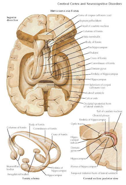
FORNIX
The fornix is an almost circular arrangement of white fibers conveying the great majority of the hippocampal efferents to the hypothalamus and carrying commissural fibers to the opposite hippocampus and habenular trigone. The fornix rises out of the fimbria of the hippocampus, which turns upward beneath the splenium of the corpus callosum and above the thalamus to form the crura (posterior columns) of the fornix. Anterior to the commissure of the fornix, the two crura unite for a variable distance in the midline and create the triangular body of the fornix. The free lateral edges of the fornix help to bind the choroid fissure, through which the pia mater of the telachoroidea becomes invaginated into the lateral ventricles.
Above the interventricular foramina,
the two halves of the body of the fornix
separate to become the (anterior) columns of the fornix. As each column
descends, it sinks into the corresponding lateral wall of the third ventricle;
the majority of its fibers end in the mammillary body, although some
also pass to other hypothalamic nuclei.
 |
| Plate 2-10 |
The fornix is the main efferent
pathway from the hippocampus to the hypothalamus. Fibers ending in the
mammillary body form synapses around its cells. The axons of these cells pass
upward in the mammillothalamic tract to the homolateral anterior thalamic
nucleus, from which they are relayed to the cingulate gyrus.
Other Structures. The habenular trigone is a small area
found bilaterally between the posterior end of the thalamus, the superior
(cranial) colliculus and the stalk of the pineal gland. Each trigone overlies a
habenular nucleus, which receives afferent fibers via the stria
medullaris of the thalamus (stria habenularis), a fine strand demarcating the superior and medial surfaces of
the thalamus. This stria conveys fibers from the anterior perforated substance,
the paraterminal gyrus and sub- callosal area, and perhaps other fibers
detached from the stria terminalis near the interventricular foramen. Most of
these fibers end in the homolateral habenular nucleus, but some decussate in
the small habenular commissure lying above the stalk of the pineal gland.
The fresh relay of fibers arising in
the habenular nucleus passes by way of the fasciculus retroflexus to the interpeduncular
nucleus in the posterior (interpeduncular) perforated substance. Efferent
fibers from the interpeduncular nucleus then descend in or near the medial
longitudinal fasciculus to be distributed to tegmental and reticular nuclei in
the brainstem. The amygdaloid body is described in Plate 2-11.


