Muscles of Pharynx Anatomy
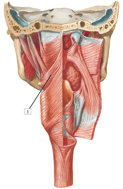
1.
Superior pharyngeal constrictor muscle
Origin:
This
broad muscle arises from the pterygoid hamulus, pterygomandibular raphe,
posterior portion of the mylohyoid line of the mandible, and side of the
tongue.
Insertion: The muscles from each side meet and attach to the median raphe of the pharynx and pharyngeal tubercle of the occipital bone.
Action:
Constricts
the wall of the upper pharynx during swallowing.
Innervation:
Pharyngeal
plexus of the vagus nerve (CN X).
Comment:
The
3 pharyngeal constrictors help move food down the pharynx and into the
esophagus. To accomplish this, these muscles contract serially from superior to
inferior to move a bolus of food from the oropharynx and laryngopharynx into
the proximal esophagus.
The
superior constrictor lies largely behind the mandible.
Clinical:
While
the motor innervation of the pharyngeal constrictors is
via the vagus nerve (CN X), the sensory innervation of all but the most
superior part of the pharynx (the constrictor muscles and the mucosa lining the
interior of the pharynx) is via the glossopharyngeal nerve (CN IX). Together,
the fibers of CN IX and X form the pharyngeal plexus and function
in concert with one another during swallowing.
1.
Middle pharyngeal constrictor muscle
Origin:
Arises
from the stylohyoid ligament and the greater and lesser horns of
the hyoid bone.
Insertion:
The
muscles from both sides wrap around and meet to attach to the median raphe of
the pharynx.
Action:
Constricts
the wall of the pharynx during swallowing.
Innervation:
Pharyngeal
plexus of the vagus nerve (CN X).
Comment:
The
middle pharyngeal constrictor lies largely behind the hyoid bone. The fibers of
the superior and middle pharyngeal constrictors often blend together, but the
demarcation point can be seen where the stylopharyngeus muscle
intervenes.
Clinical:
While
the motor innervation of the pharyngeal constrictors is
via the vagus nerve (CN X), the sensory innervation of all but the most
superior part of the pharynx (the constrictor muscles and the mucosa lining the
interior of the pharynx) is via the glossopharyngeal nerve (CN IX). Together,
the fibers of CN IX and X form the pharyngeal plexus and function
in concert with one another during swallowing.
1.
Inferior pharyngeal constrictor muscle
Origin:
Arises
from the oblique line of the thyroid cartilage and side of
the cricoid cartilage.
Insertion:
The
2 inferior pharyngeal constrictor muscles wrap posteriorly to meet and attach
to the median raphe of the pharynx.
Action:
Constricts
the wall of the lower pharynx during swallowing.
Innervation:
Pharyngeal
plexus of the vagus nerve (CN X).
Comment:
The
inferior pharyngeal constrictor lies largely behind the thyroid and cricoid
cartilages. Its lower end is referred to as the cricopharyngeal muscle, which
is continuous with the esophageal muscle fibers.
Where
the inferior constrictor attaches to the cricoid cartilage represents
the narrowest portion of the pharynx.
Clinical:
While
the motor innervation of the pharyngeal constrictors is
via the vagus nerve (CN X), the sensory innervation of all but the most
superior part of the pharynx (the constrictor muscles and the mucosa lining the
interior of the pharynx) is via the glossopharyngeal nerve (CN IX). Together,
the fibers of CN IX and X form the pharyngeal plexus and function in concert
with one another during swallowing. Injury to the pharyngeal fibers from CN X
can result in difficulty swallowing (dysphagia).
1.
Stylopharyngeus muscle
Origin:
Arises
from the styloid process of the temporal bone.
Insertion:
Attaches
to the posterior and superior margins of the thyroid cartilage.
Action:
Elevates
the pharynx and larynx during swallowing and speaking.
Innervation:
Glossopharyngeal
nerve (CN IX).
Comment:
This
muscle passes between the superior and middle pharyngeal constrictors. The
stylopharyngeus is 1 of 3 muscles arising from the styloid process of the
temporal bone (the others are the styloglossus and stylohyoid). Each muscle is
innervated by a different cranial nerve and arises from a different embryonic
branchial arch.
The
stylopharyngeus arises embryologically from the 3rd pharyngeal (branchial) arch
and is the only muscle innervated by the glossopharyngeal
nerve.
Clinical:
A
lesion to the motor fibers of CN IX that innervate the stylopharyngeus muscle
can cause pain when the patient initiates swallowing.


