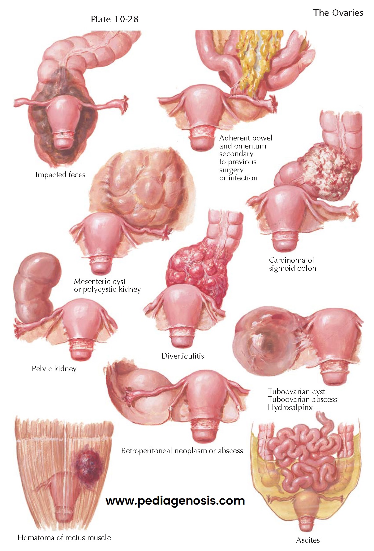
CONDITIONS SIMULATING OVARIAN NEOPLASMS II
Mesenteric
cyst. Rarely, a cyst of the mesentery of the transverse
colon or small bowel or of the omentum may reach large proportions and simulate
a pedunculated, freely movable, extrapelvic ovarian cyst.
Polycystic kidney. The multilocular cystic replacement of the renal cortex and medulla may reach a huge size. On abdominal palpation the possibility of a large, adherent ovarian cyst may be suggested. Intravenous pyelography may demonstrate the bilateral renal involvement and characteristic elongation and spreading apart of the calyces. Abdominal ultrasonography will demonstrate the classic cystic nature of the kidneys and cysts, if present in the liver.
Pelvic
kidney. An ectopic kidney may lie low in the lumbar
region, in the iliac fossa, or in the true pelvis. It may be symptomless or may
give rise to sacroiliac backache and pain in the lower abdomen, radiating to
the hips and thighs. Pelvic examination may reveal the lower end of a smooth,
oval, retroperitoneal, fixed mass with a distinctive rubbery consistency.
Retroperitoneal
pelvic neoplasms. A variety of retroperitoneal
tumors may be present in the pelvis and be mistaken for adherent ovarian
neoplasms. They may be symptomless or associated with local or referred pain
due to renal compression. On rectal or rectovaginal examination, a fixed,
retroperitoneal tumor may be felt behind or lateral to the rectum.
Sigmoidoscopy and barium enema may reveal external compression. Retroperitoneal
pelvic tumors include lipoma, fibroma, sarcoma, dermoid, malignant teratoma,
metastatic carcinoma, osteochondroma, and ganglioneuroma. Pyogenic infections
of the sacroiliac joint, osteomyelitis of the pelvis, dissecting abscesses
originating with tuberculosis of the spine, perivesical infections, and psoas
abscesses must be taken into consideration.
Hematoma
of the rectus muscle. As a result of direct trauma or
unusual strain upon the recti muscles of the abdominal wall, rupture of the
muscle fibers, with a hematoma, may occur. If localized over the right or left
lower quadrants, the tender tumescence and voluntary spasm may suggest an acute
accident in an ovarian tumor. An ecchymosis may or may not be apparent. Its
superficial location may be demonstrated by tensing the abdominal muscles. The
absence of palpable tumors, upon rectal or vaginal examination, will further
clarify the issue.
Adherent
bowel or omentum. Following pelvic surgery or as
an aftermath of pelvic infections, the omentum, sigmoid colon, or small bowel
may become adherent to one adnexa or the other. An irregular, matted mass may
result and give the impression of a pelvic tumor.
Carcinoma
of the sigmoid colon. The irregular, rather firm and
often fixed mass felt with carcinoma of the sigmoid may suggest a carcinoma of
the ovary and vice versa. Whichever site of origin is suspected, further
differentiation is indicated. Altered bowel habits, diarrhea, constipation,
colicky pain, ribbon stools, and melena point to possible intestinal difficulty.
Rectal examination, sigmoidoscopy, barium enema, and biopsy will demonstrate
the presence of a lesion or filling defect.
Diverticulitis.
Uncomplicated diverticulosis of the descending and
sigmoid colon is frequently asymptomatic. Perforation may result in a localized
abscess, the adherence of bowel, omentum, and adjacent viscera, fistulous
communications, granulomas, and stenosis of the bowel. A diverticulitis of the
sigmoid may simulate a carcinoma of the sigmoid or ovary as well as pelvic
infection.
Tuboovarian
inflammatory masses. A large hydrosalpinx or
tuboovarian cyst may be palpated as a thin-walled, retort-shaped, cystic
structure, adherent to the uterus, broad ligament, and pelvic peritoneum.
Ascites. Ascitic fluid may give the impression of a large, flaccid cyst. The percussion note over an ovarian cyst is flat, with tympany in the flanks. On bimanual examination, fluctuation may be elicited. In the presence of ascites, tympany may emanate centrally and shifting dullness may be registered in the flanks. A fluid wave may be transmitted.


