Proprioception And
Reflexes
We are aware of the orientation of our
limbs with respect to one another,
we can perceive the movements of our joints and we can accurately assess the amount
of resistance (force) that opposes the movements we make. This ability is
called proprioception. The three qualities of this modality are position,
movement and force. The receptors or proprioceptors that mediate
this modality are principally found in the joint capsules (joint receptors),
muscles (muscle spindles) and tendons (Golgi tendon organs).
The joint capsule is compressed
or stretched when the joint moves, and mechanoreceptors within it signal
the position of the joint, as well as the direction and velocity of the movement.
Individual receptors respond to the position of the joint, as well as the direction
and the velocity of the movement, but not the force. The receptor types found in
the joint capsule are Ruffini-type (slowly adapting) stretch receptors (Chapter
55).
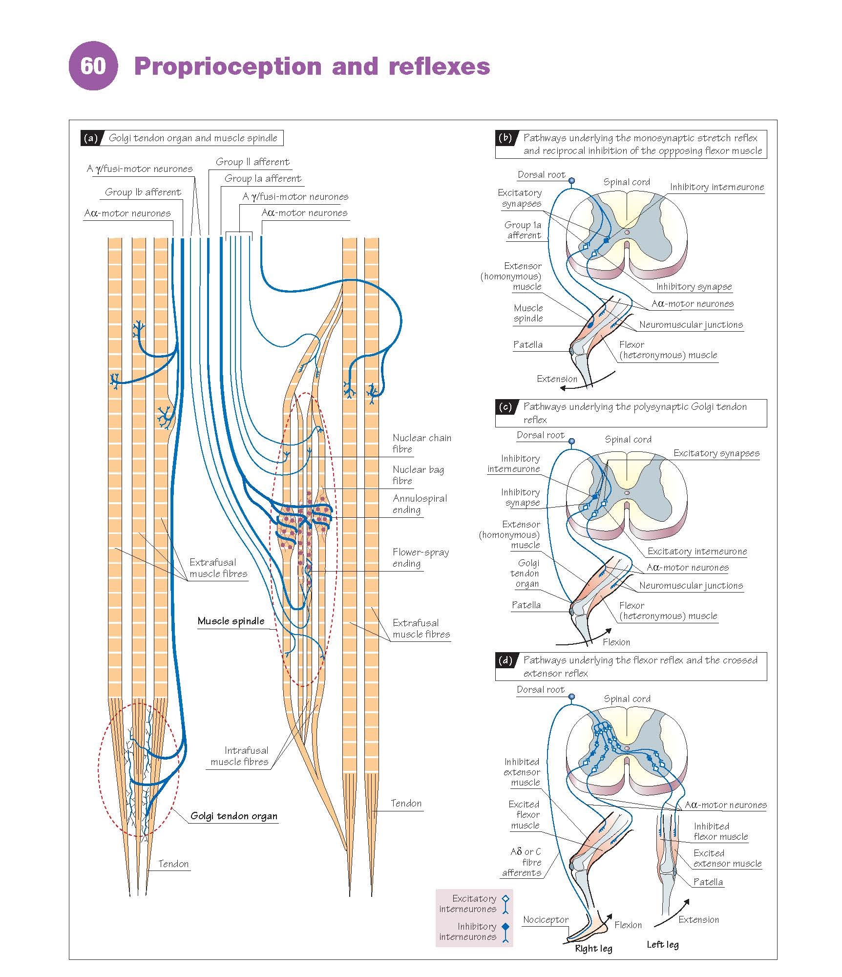
Each muscle contains a number of small
muscle fibres (intrafusal muscle fibres: 15–30 μm in diameter and 4–7 mm
in length) that are thinner and shorter than the ordinary muscle fibre (extrafusal
muscle fibres: 50–100 μm in diameter and varying in length from a few millimetres
to many centimetres). Several intrafusal fibres are grouped together and encased
in a connective tissue capsule, called the muscle spindle, a specialized
receptor that responds to the stretch of a muscle (Fig. 60a). Muscle spindles lie
in parallel to the extrafusal muscle fibres and are elongated when the muscle
is stretched. The primary sensory innervation of the muscle spindle consists of
afferent fibres which wind themselves around the centre of the intrafusal
muscle fibres (annulospiral ending). These are large myelinated fibres (group
Ia afferents). These endings are called primary sensory endings and, when
excited, they evoke a monosynaptic stretch reflex involving an excitation
of the homonymous α-motor neurones and reciprocal inhibition of the heteronymous
α-motor neurones (Fig. 60b).
Many muscle spindles also have a secondary
sensory innervation (group II afferent). They are thinner and end in flower-spray
endings (they are not involved in the monosynaptic stretch reflex).
The intrafusal muscle fibres possess
a motor innervation, the Aγ- or fusi-motor neurones. They are smaller in diameter
than those innervating the extrafusal muscle fibres, the Aα-motor neurones.
Stretching the muscle, thereby extending
both the extrafusal and intrafusal muscle fibres, can excite the muscle spindle.
However, there is a second way to excite the primary muscle spindle ending – by
contraction of the intrafusal muscle fibre brought about by excitation of the fusi-
or γ-motor neurone. This does not change the overall length or tension of the entire
muscle, as it is too weak; however, it is sufficient to stretch the central portion
of the intrafusal fibres, inducing excitation in the primary sensory ending.
Contraction of the extrafusal fibres
can be triggered or at least facilitated
by the muscle spindle, either by stretch of the whole muscle or by activation of
the fusi- or γ-motor neurones. They can comple- ment each other or have a mutually
cancelling effect. The threshold of the stretch reflex can also be varied by intrafusal
activity.
The Golgi tendon organs are stretch
receptors found in the muscle tendons (Fig. 60a). Each receptor is associated with
the tendon fascicle of about 10 extrafusal muscle fibres, is surrounded by a capsule
of connective tissue and is innervated by large myelinated afferent fibres (group
Ib fibres). They are in series with the extrafusal muscle fibres and respond
to tension in the muscle. They can respond both when the muscle contracts and when
the muscle is stretched, unlike the muscle spindle which responds predominantly
during stretching of the muscle. Golgi tendon organs protect against overloading
and provide a protective reflex. In a functional sense, the segmental connections
of the Ib fibres are a mirror image of the Ia fibres. A marked increase in muscle
tension, whether resulting from stretch, contraction or a combination of the two,
will result in inhibitory connections with the homony- mous motor
neurones and excitatory connections with the heteronymous motor neurones.
However, none of these connections is monosynaptic; all involve at least two synapses
(Fig. 60c).
The properties of the joint receptors
make it very likely that they are primarily responsible for mediating the sense
of position and movement. The most likely detectors of force sensation are the muscle
spindles and Golgi tendon organs. Other receptors also contribute to the sense of
force, as well as movement and position, such as mechanoreceptors in the skin.
Polysynaptic motor reflexes. Many receptors in the body, other than those found
in muscle, can trigger motor reflexes. Experiments on animals, in which the spinal
cord has been severed, have shown that many of these reflexes are restricted to
the spinal cord. They are always polysynaptic. The most prominent examples of these
reflexes are the flexor reflex and the crossed extensor reflex (Fig. 60d). If a
pinprick is made to a toe, the stimulated limb is pulled away. The flexor reflex
is a flexion of the knee and hip joints. It is a protective reflex, pulling
the limb from the site of the noxious stimulus. The delay and the magnitude of the
response are very much dependent on the stimulus intensity. The higher the intensity,
the shorter the latency and the quicker the response. It can also be observed that
the flexion of one limb is always accompanied by the extension of the limb on the
other side of the body. In other words, there is an ipsilateral flexor reflex and
a contralateral extensor reflex. This contralateral extensor reflex is called the
crossed extensor reflex. These reflexes are not only enhanced in spinal animals,
but also in newborn and premature babies, as during the days just after birth the
higher levels of the brain are not fully
developed.


