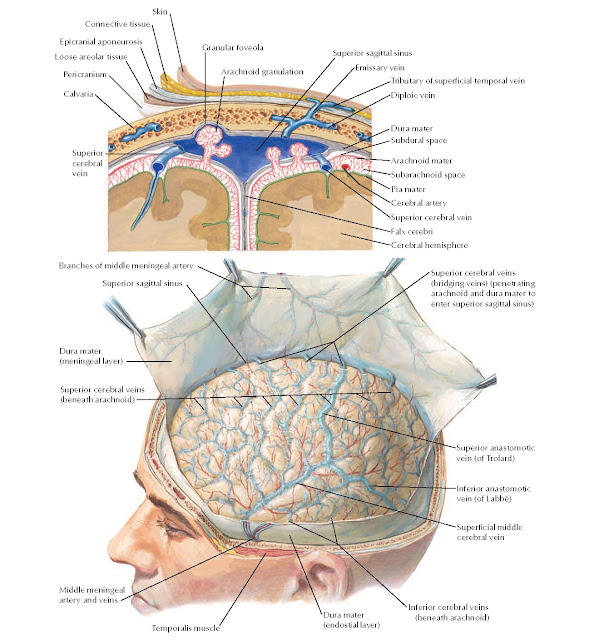
Meninges and Superficial Cerebral Veins Anatomy
Epicranial aponeurosis, Loose areolar tissue, Skin, Connective tissue, Arachnoid granulation, Superior sagittal sinus, Emissary vein, Tributary of superficial temporal vein, Diploic vein, Dura mater, Arachnoid mater, Subarachnoid space, Pia mater, Cerebral artery, Superior cerebral vein, Falx cerebri, Cerebral hemisphere, Dura mater (meningeal layer),
Branches of middle meningeal artery, Superior sagittal sinus, Pericranium, Calvaria, Granular foveolar, Superior anastomotic vein (of Trolard), Inferior
anastomotic vein (of
Labbé), Superficial middle
cerebral vein, Middle meningeal artery and veins, Temporalis muscle, Dura mater
(endostial layer), Superior cerebral veins (beneath arachnoid), Superior cerebral veins (bridging veins) (penetrating arachnoid and dura mater to enter superior sagittal sinus), Subdural space, Inferior
cerebral veins (beneath
arachnoid), Superior
cerebral vein.


