Masseter, Temporalis and Infratemporal Fossa
Masseter
(Fig. 7.29) attaches along the length of the
zygomatic arch and its fibres slope downwards and backwards to the lateral
surface of the ramus of the mandible adjacent to the angle (Fig. 7.31). This
muscle is a powerful elevator of the mandible
and is easily palpated when the teeth are clenched. It is supplied by the
masseteric branch of the mandibular (V3) division of the trigeminal nerve.
Temporalis
Temporalis
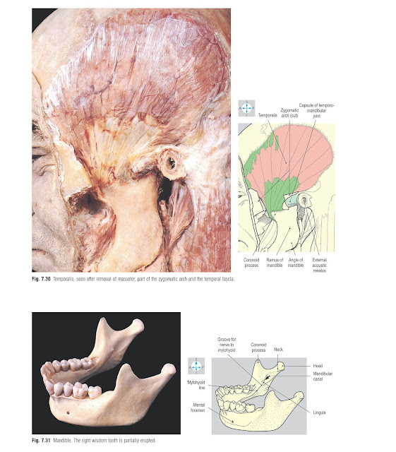
(Fig. 7.30) is a large fan-shaped muscle occupying the temporal fossa and
taking attachment from the area of bone bounded by the inferior temporal line.
The more superficial fibres arise from the temporal fascia that covers the
muscle and is attached to the superior temporal line. All the fibres descend
deep to the zygomatic arch to attach to the coronoid process and anteromedial
aspect of the ramus of the mandible (Fig. 7.31). Temporalis elevates the
mandible, as in closing the mouth, and its posterior fibres retract the
mandible. The deep temporal branches of the mandibular (V3) division of the
trigeminal nerve supply the muscle from its deep surface.
Infratemporal
fossa
This
fossa lies deep to the ramus of the mandible and is limited on its medial
aspect by the lateral wall of the pharynx and the medial pterygoid plate of the
sphenoid bone. The fossa is bounded by the posterior surface of the maxilla in
front and by the styloid process and its attached muscles behind. The roof is
provided by the temporal and sphenoid bones in the base of the skull while
inferiorly the fossa is continuous with the neck.
Within
the fossa are the two pterygoid muscles, the mandibular (V3) division of the
trigeminal nerve and its branches, and the maxillary vessels and their
branches. Adjacent to the fossa is the temporomandibular joint.
Pterygoid
muscles
Each
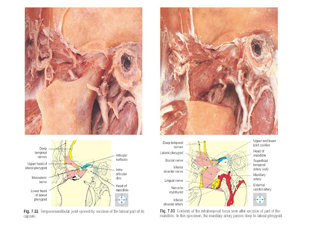
of the lateral and medial pterygoid muscles (Figs 7.32–7.34) has two
attachments to the skull. The upper head of the lateral pterygoid attaches to
the inferior surface of the greater wing of the sphenoid. The lower head
attaches to the lateral surface of the lateral pterygoid plate. Both heads
converge on the neck of the mandible and the capsule of the temporomandibular
joint. The lateral pterygoid pulls forward both the neck of the mandible and
the articular disc, thus depressing the mandible and opening the mouth.
The
lower head of the lateral pterygoid is clasped by the two heads of the medial
pterygoid. The deep head of the latter is larger and attaches to the medial
surface of the lateral pterygoid plate. The superficial head is attached to the
tuberosity of the maxilla. The fibres of both heads incline obliquely
downwards, backwards and laterally to attach to the medial surface of the angle
of the mandible. The muscle is a powerful elevator of the mandible.
The
temporomandibular joint (Fig. 7.32) is a
synovial joint. The head of the mandible articulates with the articular fossa
and eminence of the temporal bone. Fibrocartilage covers the articular surfaces
and also forms an articular disc, which divides the joint into two separate
cavities. Within these cavities, the non-cartilaginous surfaces are lined with
synovial membrane.
The
fibrous capsule surrounding the joint is attached to the margin of the
articular cartilage and to the neck of the mandible. Anteriorly, it receives
the attachment of the lateral pterygoid while its deep surface is firmly
adherent to the periphery of the articular disc.
Laterally,
the capsule (Fig. 7.30) is thickened to form the lateral ligament, which
inclines posteroinferiorly from the root of the zygomatic arch to the neck of
the mandible. Two accessory ligaments lie medial to the joint, although not in
contact with the capsule. The sphenomandibular ligament extends from the spine
of the sphenoid to the lingula adjacent to the mandibular foramen. The
stylomandibular ligament, a thickening of the parotid fascia, passes from the
styloid process to the angle of the mandible.
The
joint receives its nerve supply from the auriculotemporal and masseteric
branches of the mandibular (V3) division of the trigeminal nerve.
Movements
at the joint include elevation, depression, protraction and retraction of the
mandible. The head of the mandible does not merely rotate in the articular
fossa but also moves forwards onto the articular eminence of the temporal bone,
taking the articular disc with it. The alternate protraction and retraction of
right and left sides produces the grinding movements used in chewing. The
muscles responsible for these movements are known collectively as the muscles
of mastication. The mouth is closed by contraction of masseter, temporalis and
medial pterygoid. The lateral pterygoid protracts the mandible and, assisted by
digastric and mylohyoid (p. 348), also opens the mouth. Retraction is produced
by the posterior fibres of temporalis. When the mandible is fully depressed,
the joint is relatively unstable and dislocation may occur, the head of the
mandible moving in front of the articular eminence and resulting in an
inability to close the mouth.
The
mandibular division of the trigeminal nerve (Figs 7.33 & 7.34) contains
sensory and motor fibres and enters the infratemporal fossa through the foramen
ovale in the sphenoid. Two small branches arise from the short main trunk of
the nerve. The first branch ascends through the foramen spinosum to receive
sensation from the meninges of the middle cranial fossa. The other branch is
motor, supplying the medial pterygoid and giving a small branch that passes
through the otic ganglion (lying just medial to the main trunk of the
mandibular division) to supply tensor tympani and tensor veli palatini.
The
main trunk descends between the lateral pterygoid and tensor veli palatini
muscles, dividing into anterior and posterior divisions. The anterior division
is mainly motor and gives masseteric, deep temporal, lateral pterygoid and
buccal branches. The masseteric nerve (Fig. 7.32) curves laterally above the
lateral pterygoid to enter the deep surface of masseter. Two or three deep
temporal nerves (Fig. 7.33) ascend deep to temporalis, which they supply, and
further branches enter the deep surface of the lateral pterygoid. The buccal nerve
(Figs 7.33 & 7.34) is a sensory branch that passes forwards between the two
heads of the lateral pterygoid to supply the skin over the cheek and the mucosa
lining the cheek, which it reaches by piercing, but not supplying, buccinator.
The
posterior division of the main trunk is mainly sensory and has three branches,
the auriculotemporal, lingual and inferior alveolar nerves. The
auriculotemporal nerve (Fig. 7.34) arises
by two roots, which clasp the origin of the middle meningeal artery. The nerve
passes backwards before turning superiorly behind the temporomandibular joint
to ascend in company with the superfi- cial temporal vessels. It gives
secretomotor branches to the parotid gland (p. 339) and conveys sensation from
the temporal region, the upper half of the pinna and most of the external
acoustic meatus.
The
lingual nerve (Figs 7.33 & 7.34) inclines downwards and forwards between
the pterygoids, deviating medially to pass below the superior constrictor of
the pharynx. In the floor of the mouth it runs forwards lateral to the
hyoglossus muscle, at whose anterior border it again turns medially to pass
inferior to the submandibular duct and enter the base of the tongue. It conveys
general sensation from the anterior two-thirds of the tongue. Near the lower
border of the lateral pterygoid the lingual nerve is joined by the chorda
tympani (a branch of the facial nerve). Arising within the temporal bone, the
chorda tympani emerges from the petrotympanic fissure. It carries taste fibres,
which have travelled in the lingual nerve from the anterior two-thirds of the
tongue and preganglionic parasympathetic fibres destined for the submandibular
ganglion (p. 352).
The
inferior alveolar nerve (Figs 7.33 & 7.34) descends medial to the lateral
pterygoid and gives rise to a motor branch that curves downwards to supply
mylohyoid and the anterior belly of digastric. The inferior alveolar nerve then
enters the mandibular foramen in the ramus of the mandible and runs forwards in
the mandibular canal, supplying the lower teeth and alveolar ridge. Its mental
branch emerges from the mental foramen to supply skin overlying the chin. Local
anaesthetic injected near the inferior alveolar nerve as it enters the
mandibular foramen will block sensation from the lower teeth and gums on that
side of the mouth. Often there is loss of sensation in the same side of the
tongue because of the proximity of the lingual nerve
This
artery (Figs 7.33 & 7.34) arises in the parotid gland (p. 339) as a
terminal branch of the external carotid artery, passes antero- superiorly
across the infratemporal fossa, usually lateral to the lateral pterygoid, and traverses the
pterygomaxillary fissure to enter the pterygopalatine fossa where terminal branches
arise. These correspond to branches of the maxillary nerve (p. 353).
In
the infratemporal fossa, the maxillary artery gives branches to supply
masseter, temporalis and the pterygoid muscles. In addition, the middle
meningeal artery arises deep to the lateral pterygoid, embraced by the two
roots of the auriculotemporal nerve. It traverses the foramen spinosum and
within the cranium supplies the meninges of the middle cranial fossa and the
cranial vault.
The
maxillary artery also gives rise to the inferior alveolar artery, which
accompanies the nerve into the mandibular canal. Further small branches supply
the middle ear and the lining of the external acoustic meatus.
Maxillary artery
Veins
within the pterygopalatine fossa form a plexus that extends through the
pterygomaxillary fissure into the infratemporal fossa, where the plexus is
related to the pterygoid muscles. This pterygoid plexus has important
connections to the cavernous sinus in the skull and infraorbital and ophthalmic
veins. The plexus drains by the maxillary vein into the retromandibular vein
(p. 340).


