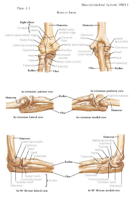
ELBOW JOINT
The elbow
joint, comprising the humeroradial, humeroulnar, and proximal radioulnar
articulations within a common capsule, necessarily involves the proximal
portions of the radius and ulna, as well as the distal part of the humerus. The
humeroulnar articulation acts as a hinge and allows flexion and extension of
the elbow, whereas rotational movements occur through the humeroradial and
proximal radioulnar articulations. Therefore, the elbow is not considered a
simple hinge joint but rather a trochoginglymoid joint that possesses two
degrees of freedom or motion: flexion-extension and pronation-supination. The
center of rotation of the elbow runs through the center of the articular
surface of the distal humerus formed by the trochlea and the capitellum, lying
just anterior to the anterior cortex of the distal humerus on the lateral view.
The carrying angle of the elbow, formed by the humerus and ulna with the hand
and forearm fully supinated and the elbow fully extended, has been reported to
range from 11 to 14 degrees of valgus in men and from 13 to 16 degrees of
valgus in women. The carrying angle is typically about a degree greater on the
dominant compared with the nondominant side. Valgus (cubitus valgus) or varus
(cubitus varus) malalignment is diagnosed when the carrying angle is greater
than or less than these normal values, respectively.
In the region of the elbow joint, the
special parts of the radius are the head, neck, and tuberosity. The radial head
is round; it is a thick disc, articular both on its circumference and over its
cupped upper free surface. The latter surface articulates with the capitellum
of the humerus. The articular circumference of the head is broader medially for
contact with the radial notch of the ulna, forming a 240-degree arc for
articulation with the ulna, and narrower where it is held by the annular
ligament. The neck of the radius is the constriction below the head, and the
tuberosity is an oval prominence just distal to the neck. Its posterior portion
is roughened for the reception of the biceps brachii tendon; its anterior part
is smooth and is in contact with the bicipitoradial bursa.
The proximal end of the ulna has a
more complex architecture. Its heavy proximal extremity exhibits the opened
jaws of the trochlear notch, the olecranon, the coronoid process, and the
radial notch.
The trochlear
notch is a concavity that describes about one third of a circle and is
divided by a longitudinal ridge into medial and lateral parts. The waist of the
notch is constricted, and a roughness across the waist separates the part
deriving from the olecranon from the part formed by the coronoid process. The
trochlear notch receives the trochlea of the humerus. In extreme flexion, the
coronoid process of the ulna enters the coronoid fossa of the humerus; in
extreme extension, the olecranon enters the olecranon fossa.
The olecranon contributes part
of the trochlear notch and forms the posterior projection of the elbow. Its
blunt end receives the tendon of the triceps brachii muscle and is attached to
the capsule of the elbow joint along the bounding margin of the trochlear
notch. Between these attachments, the bone is smooth for the subtendinous bursa
of the triceps brachii muscle.
The coronoid process is a
strong, triangular projection of the anterior surface of the ulna; it also
forms the anterior part of the trochlear notch. The anterior surface of the
coronoid process is rough for the insertion of the tendon of the brachialis
muscle. The junction of this surface with the shaft is the location of the
tuberosity of the ulna, which receives the oblique cord of the radius.
The radial notch of the ulna, a
shallow concavity on the lateral aspect of the coronoid process, receives the circumferential
articular surface of the head of the radius. Its prominent edges give
attachment to the ends of the annular ligament of the radius.
Ossification
The radius is ossified by means of a
center of ossifica- tion that appears in the shaft (which is bony at birth) at the
eighth week of intrauterine life. Ossification for the radial head begins at 4
or 5 years of age, and this portion fuses with the shaft at 14 to 16 years of
age.
Ossification of the ulna begins near
the middle of the shaft at the eighth w ek of intrauterine life, and the shaft
is bony at birth.


