FRACTURE OF HEAD AND
NECK OF RADIUS
Fractures of the radial head occur primarily in adults, whereas fractures
of the radial neck are more common in children. The usual causes of these
injuries are indirect trauma, such as a fall on the outstretched hand, and,
less commonly, a direct blow to the elbow. Radial head and neck fractures are
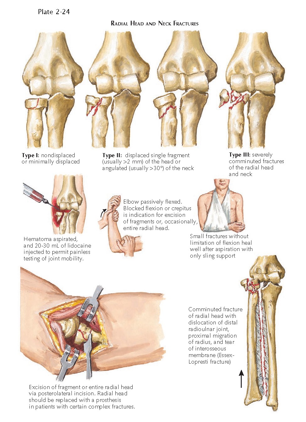
generally classified into four groups. In type I fractures, the fracture is
nondisplaced or minimally displaced. Type II fractures refer to displaced
fractures of the joint margin or neck with a single fracture line. Type III
fractures are comminuted fractures of the head or neck. Type IV fractures are
associated with dislocation of the elbow.
Diagnosis of a radial or neck head
fracture may be difficult. Pain, effusion in the elbow, and tenderness to
palpation directly over the radial head or neck are the typical manifestations.
If the fracture is displaced, a “click” or crepitus over the radial head or
neck is detected during forearm supination or pronation. Radiographic findings
in nondisplaced fractures are minimal, and the radiograph often shows only
swelling in the elbow with a fat pad sign. Any radiographic evidence of fat pad
displacement accompanied by tenderness over the radial head or neck strongly
suggests a fracture.
Treatment of a radial head or neck
fracture depends on careful clinical and radiographic evaluation. Type I fractures
can be managed nonoperatively if they appear nondisplaced or minimally
displaced on radiographs and demonstrate no evidence of a mechanical block on
elbow range of motion. To determine the presence of a mechanical block, the
elbow joint can be aspirated to remove the bloody effusion, followed by
injection of lidocaine into the joint to relieve pain and allow a thorough
examination. The examiner can then move the elbow painlessly through a full
range of motion to assess the degree of flexion and extension and of pronation and
supination and to detect any crepitus or blocked motion due to a displaced
fragment. If the range of motion is adequate and there is no bone block or
significant crepitus, then the elbow is placed in a posterior splint for a few
days. After this period, the patient can remove the splint and begin active
range-of-motion exercises for the injured elbow. Frequent follow-up radiographs
are necessary to detect any late displacement of the fracture fragment.
Controversy surrounds the treatment of
displaced (type II) and comminuted (type III) fractures of the radial head or
neck and fractures associated with limited range of motion due to a fracture
fragment. Surgical fixation is indicated for fractures with one or two large,
displaced fragments that can be effectively reduced and stabilized with a plate
and/or screws or Kirschner wires. Comminuted fractures that cannot be
adequately reduced and stabilized with surgery usually require excision of the
radial head. When the radial head is removed, the annular ligament must be
preserved to maintain the integrity of the ligament complex of the proximal
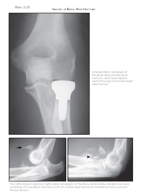
radioulnar joint. Radial head implants can be placed after radial head
excision, but care should be taken to avoid oversizing the prosthesis, which
can limit elbow range of motion. A radial head replacement should always be
used after resection of the radial head when an Essex-Lopresti injury is
present (fracture of the radial head with dislocation of the distal radioulnar
joint and disruption of the interosseous membrane). In an Essex-Lopresti
fracture, the radius will migrate proximally if the radial head is not replaced
after excision, which is very debilitating to the entire forearm complex (see
Plate 2-24). Placement of a radial head implant prevents proximal migration of
the radius and minimizes long-term complications.
Dislocations of the elbow with
comminuted fractures of the radial head or neck (type IV) are serious injuries
that usually involve significant soft tissue injury. Both the joint capsule and
the collateral ligaments of the elbow can be damaged, and the joint injury can
lead to stiffness or persistent instability, osteoarthritic changes, and
myositis ossificans. If surgery is appropriate and feasible, these type IV
injuries should be surgically repaired early or replaced to decrease the
occurrence of complications, such as stiffness or instability and myositis
ossificans. Radial head fractures can also be associated with other injuries
about the elbow, such as fractures of the capitellum, coronoid process, or
olecranon. The combined injury pattern of an elbow dislocation associated with
both a radial head and a coronoid process fracture has been termed a terrible
triad injury.


