Stomach Anatomy
The stomach is the dilated portion of the
gut, in which the early stages of digestion take place. It lies in the upper
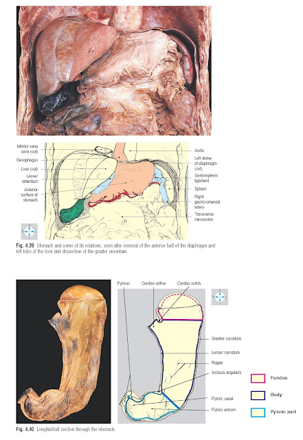
part of the abdomen beneath the left dome of the diaphragm (Fig. 4.39). Proximally, the stomach joins the oesophagus at the cardiac orifice and
distally, it is continuous with the duodenum at the pylorus. Between these two
relatively fixed points, the organ varies considerably in size, shape and
location in response to its muscle tone, the quantity and nature of its
contents and the position of the individual (Figs 4.41 & 4.42). Usually,
the loaded stomach is J-shaped and lies in the left hypochondrium, the
epigastrium and umbilical region of the abdomen.
The oesophagus pierces the diaphragm and has a short
intra-abdominal course before joining the stomach at the cardiac orifice. This
lies a little to the left of the midline at about the level of the eleventh
thoracic vertebra (Fig. 4.42). Anatomical and physiological factors produce a
sphincteric effect at the gastro-oesophageal junction. If this mechanism fails,
gastric contents can regurgitate into the oesophagus (gastro-oesophageal
reflux), causing inflammation of the oesophageal mucosa. The stomach has two
surfaces, anterior and posterior, which meet at two curved borders, the
curvatures (Fig. 4.40). The lesser curvature
extends from the cardiac orifice downwards and to the right, to reach the upper
border of the pylorus. A notch, the incisura angularis, is usually present on
the lesser curvature towards its pyloric end. The greater curvature is longer
and begins at the cardiac notch on the left side of the
cardiac orifice. It arches upwards and to the left before descending along the
left and inferior aspects of the organ to reach the inferior border of the
pylorus.
The pylorus is normally situated just to the right of the
midline at the level of the first lumbar vertebra, on the transpyloric plane.
By convention, the stomach is described as having three
parts, the fundus, the body and the pyloric part (Fig. 4.40). The fundus lies
above an imaginary horizontal plane passing through the cardiac orifice, while
the antrum lies to the right of the incisura angularis. The body lies between
the fundus and the pyloric part and is the largest part of the stomach. In the pyloric part, the
cavity of the pyloric antrum tapers to the right into a narrow passage, the
pyloric canal.
The mucosal lining presents numerous longitudinal folds
or rugae, which are most prominent when the stomach is empty (Fig. 4.40). There
is a well-developed smooth muscle coat, which is thickened around the pyloric
canal and pylorus to form the pyloric sphincter.
Relations
The anterior surface of the stomach lies in contact with
the diaphragm, the anterior abdominal wall and the left and quadrate lobes of
the liver. Posterolateral to the fundus lies the gastric surface of the spleen
(Fig. 4.39). The remainder of the stomach’s relations are situated posteriorly
and collectively form the stomach bed. This includes the diaphragm, left
suprarenal gland, upper part of the left kidney, the splenic artery, pancreas,
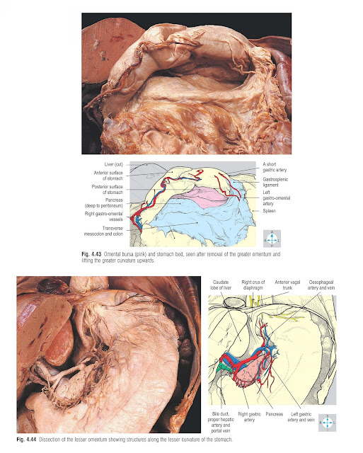
transverse mesocolon and sometimes, the transverse colon (Fig. 4.43). However,
these structures are separated from the stomach by the omental bursa (p. 157).
Gastric ulcers can perforate into either the greater sac or the omental bursa.
Sometimes, ulceration may involve the pancreas or the splenic artery.
Attached to each curvature of the stomach is an omentum,
a double layer of peritoneum. The lesser omentum extends from the liver (Fig.
4.37) to the lesser curvature and also attaches to the abdominal oesophagus and
the commencement of the duodenum (Fig. 4.39). Near the lesser curvature, this
omentum contains the left and right gastric vessels (Fig. 4.44), accompanied by lymphatics and autonomic nerves, while its free border
encloses the portal vein, the bile duct and the proper hepatic artery.
The greater omentum hangs from the distal part of the
greater curvature and from the superior duodenum. Near the greater curvature,
it contains the left and right gastro-omental (gastroepiploic) vessels (Fig. 4.43). To the left, the greater omentum is continuous with the gastro-splenic
ligament, which connects the proximal part of the greater curvature to the
hilum of the spleen.
The stomach is supplied by several arteries, which are
all derived from branches of the coeliac trunk and which anastomose extensively with each other. The coeliac trunk (Fig. 4.45) is a short, wide vessel arising from the anterior aspect of the aorta
just below the diaphragm. It divides into three branches: the left gastric, the
common hepatic and the splenic arteries.
The left gastric artery is the smallest branch, passing
upwards and to the left behind the omental bursa to reach the oesophagus, then
descending along the lesser curvature within the lesser omentum (Fig. 4.44).
Its branches include two or three to the lower oesophagus, which ascend through
the oesophageal opening of the diaphragm. Other branches supply the cardia and
lesser curvature of the stomach.
The common hepatic artery gives rise to the right gastric
and gastroduodenal arteries. The right gastric artery (Fig. 4.44) arises above
the superior duodenum and runs to the left within the lesser omentum, supplying
the lesser curvature and anastomosing with the left gastric artery. One of the
branches of the gastroduodenal artery is the right gastro-omental (gastroepiploic) artery (Fig. 4.43). This vessel runs to the left within the greater
omentum, parallel to the greater curvature, giving numerous branches to the
pyloric part and body of the stomach.
The splenic artery is the largest branch of the coeliac
trunk (Fig. 4.45). It runs a tortuous
course to the left along the superior border of the pancreas, initially behind
the omental bursa and then within the splenorenal ligament, and terminates near
the hilum of the spleen. It provides collateral branches to the pancreas and
terminal branches to the spleen and stomach. There are several gastric
branches, which pass to the greater curvature by way of the gastrosplenic
ligament. Most of these vessels supply the fundus of the stomach and are called
short gastric arteries (Fig. 4.43). However, one branch, the left
gastro-omental (gastroepiploic) artery, continues downwards and to the right
within the greater omentum. It follows the greater curvature, supplies the body
of the stomach and may anastomose with the right gastro-omental
(gastroepiploic) artery.
Fig. 4.45 Stomach and most of the pancreas have been
removed to reveal the coeliac trunk and its branches.
Venous drainage
The veins of the stomach accompany the gastric arteries
and drain into the portal venous system, the portal vein itself receiving the
right and left gastric veins. The splenic vein receives the short gastric and
left gastro-omental (gastroepiploic) veins, while the right gastro-omental vein
usually enters the superior mesenteric vein. The oesophageal tributaries of the
left gastric vein (Fig. 4.44) take part in an important portacaval anastomosis
(p. 185) with tributaries of the azygos venous system within the thorax.
Nerve supply
In the thorax, the vagus nerves form a plexus on the
surface of the oesophagus. From this plexus emerge two principal nerves, the
anterior and posterior vagal trunks, which enter the abdomen on the respective
surfaces of the oesophagus. The anterior vagal trunk (Fig. 4.44), derived
mostly from the left vagus nerve, gives branches to the anterior surface of the
stomach, including the pyloric region. Branches from the posterior trunk, whose
origin is mainly from the right vagus nerve, pass to the posterior surface of
the stomach and also to the coeliac plexus (pp 197, 199). The parasympathetic
innervation of the stomach by the vagus nerves is important in relation to both
secretion and motility of the organ.
Get more articles about sciences, anatomy, physiology, abdomen here


