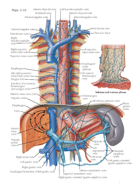
Venous Drainage of Esophagus
The venous drainage of the esophagus is effected by tributaries that
empty into the azygos and hemiazygos systems. Drainage begins in a submucosal
venous plexus, branches of which, after piercing the muscle layers, form a
venous plexus on the external surface of the esophagus. Tributaries from the
cervical esophageal veins drain into the inferior thyroid vein, which
empties into the right or left brachiocephalic vein, or into both.
Tributaries from the thoracic esophageal veins on the right side join the azygos,
right brachiocephalic, and, occasionally, vertebral vein. On the
left side they join the hemiazygos, accessory hemiazygos, left
brachiocephalic, and, occasionally, vertebral vein. Venous tributaries from
the abdominal esophagus drain mostly into the hepatic portal vein by way of the
left gastric vein, and to a lesser degree, the short gastric veins. A
small amount of venous blood from the abdominal esophagus may drain to the left
inferior phrenic vein before joining the inferior vena cava directly or via
the suprarenal and then left renal vein.
The composition and arrangement of the
azygos system of veins are extremely variable. The azygos vein arises in
the abdomen from the ascending right lumbar vein that receives the first
and second lumbar and the subcostal veins. It may arise directly from the
inferior vena cava or have connections with the right common iliac, or renal,
vein. In the thorax it receives the right posterior intercostal veins from the
fourth to the eleventh spaces and terminates by entering the right side of the
superior vena cava. The highest intercostal vein from the first space drains
into the right brachiocephalic or, occasionally, into the vertebral vein. The
veins from the second and third spaces unite in a common trunk (right
superior intercostal) that ends in the terminal arched portion of the
azygos. The hemiazygos vein arises as a continuation of the left
ascending lumbar or from the left renal vein. It receives the left
subcostal vein and the intercostal veins from the 8th or 9th to the 11th spaces
and then crosses the vertebral column posterior to the esophagus to join the
azygos. The accessory hemiazygos vein receives the intercostal veins
from the fourth to the seventh or eighth spaces and then crosses the spine
posterior to the esophagus to join the hemiazygos or to end separately in the
azygos. Above, it may communicate with the left superior intercostal that
drains the second and third spaces and ends in the left brachiocephalic.
Drainage of the first space is into the left brachiocephalic or vertebral vein.
Often the hemiazygos, accessory hemiazygos,
and superior intercostal trunk form a continuous longitudinal venous channel,
with no connections to the azygos vein on the right. On the other end of the
spectrum, the azygos vein may be located along the anterior aspect of the
vertebral body and veins from the left side of the thorax may drain directly to
it and a hemiazygos or accessory hemiazygos vein are not formed. The left azygos
system may be reduced to a slender channel, the main left venous drainage of
the esophagus and the intercostal spaces then being in the veins of the respective
vertebrae. Interruptions in the left azygos system by crossing to the right
azygos usually occur between the seventh and ninth intercostal veins, the most
common vertebral level of crossing being T8.
At the inferior end of the
esophagus, branches from the left gastric vein are continuous with the lower
esophageal branches. Portal hypertension may shunt blood
into the lower esophageal branches and there after into the superior vena cava
via the azygos and hemiazygos veins. These vessels may dilate, creating
esophageal varicosities. From this same region, blood may be shunted into the
splenic vein, retroperitoneal veins, and inferior phrenic vein of the
diaphragm, reaching the caval system. Because short gastric veins pass up from
the splenic to the cardioesophageal end of the stomach, thrombosis of the
splenic vein may redily lead to esophageal varices and fatal
hemorrhages.


