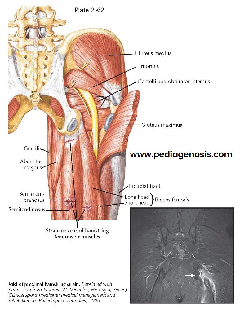
MUSCLE STRAINS
Muscle strains most commonly occur at the musculotendinous junction.
Most muscle strain injuries occur as the result of a forceful eccentric muscle
contraction. The muscles most commonly involved are those that cross two joints
(e.g., hamstrings, gastrocnemius). Another factor that makes certain muscles susceptible
to strains is an increased percentage of fast twitch muscle fibers (type II).
HAMSTRING STRAIN
The hamstring musculature (biceps femoris, semitendinosus, and semimembranosus) act to extend the hip and flex the knee. The muscle group can be injured any-where along its course from the ischium to the fibular head (biceps femoris) or medial tibia (semitendinosus, semimembranosus). Patients will often describe a pulling/tearing/ripping sensation of the posterior thigh. The most common inciting event is a change in speed while running during athletic activity.
Examination of patients with a hamstring injury
includes inspection for ecchymosis along the posterior thigh. Palpation over
the point of maximal tenderness for a defect, especially proximal, should be
undertaken. Resisted prone knee flexion strength at varying degrees and the
ability to actively extend the hip off of the examination table should be
compared.
The differential diagnosis is proximal hamstring
avulsion/sciatica.
Imaging for hamstring strains is often not necessary
when the injury occurs in the midportion of the posterior thigh. Injuries that
occur more proximal warrant plain radiographs and an MRI to evaluate for bony
avulsion or soft tissue avulsion of the proximal origin. Treatment of
midsubstance or musculotendinous injury is very similar. An initial period of
rest, ice, compression, and elevation to quell the acute phase of inflammation,
while maintaining the length of the muscle, is initiated. Once these initial
symptoms subside, isometric followed by isotonic and isokinetic exercises are
progressed until a full return to activity based on symptoms is achieved.
Recovery from a hamstring strain may range from 3 weeks to 6 months depending
on severity. Treatment of proximal avulsions may be surgical with repair back
to the ischial origin.
Proceeding to surgery is guided by factors such as
tendon displacement and patient expectations.
ADDUCTOR STRAINS
Sudden eccentric contraction, most commonly with
lateral movement and hyperabduction, is the most common mechanism of injury.
Injury events range from slips on a wet floor to those sustained during a
sporting activity, commonly soccer, hockey, and basketball. The adductor longus
muscle is the most commonly injured muscle-tendon unit.
Examination of patients with adductor strain includes
inspection for ecchymosis along the medial thigh and palpation of the adductor
origin in the pubic rami. Strength comparison and causation of pain should be
examined with the hips and knees fully extended and with both flexed to 45
degrees.
The differential diagnosis includes osteitis pubis
(athletic pubalgia, sports hernia, hip joint irritation [labral tear]).
Plain radiographs and an MRI are used to evaluate an
avulsion injury or in the case of chronic adductor issues to evaluate for partial tearing.
Groin injuries with rare exception respond to
conservative management of activity modification, gradual stretching, and
graded return to activities based on symptoms.
HIP FLEXOR STRAIN (RECTUS FEMORIS, ILIOPSOAS)
The rectus femoris is the most commonly injured muscle
of the quadriceps muscle group. It can be injured at any point along its
course. As with other strains, musculotendinous injury is most common, although
proximal injuries, especially in adolescents, must be evaluated for avulsion of
the anterior inferior iliac spine (origin). Eccentric contraction again is the
most common mechanism, especially during sprinting. The iliopsoas is less
commonly injured in isolation and may be more commonly irritated with snapping iliopsoas and iliopsoas bursitis.
Examination includes inspection for ecchymosis and
palpation of the muscle belly, observing for defects. The patient’s range of
hip motion is tested with the Thomas test while lying supine and the Ely test
lying prone, looking for hip flexion contracture and overall tight-ness.
Strength can be tested with the straight-leg raising test (rectus femoris) and
upright hip flexion (iliopsoas isolation). A strength comparison and
provocation of pain with these tests help determine the cause.
Differential diagnosis is hip joint pain (labral tear,
femoroacetabular impingement, osteoarthritis) or groin strain/sports hernia.
Standard radiographs of the pelvis are commonly
obtained to evaluate for anterior inferior iliac spine avulsion as well as for
other entities in the differential diagnosis.
The modicum of activity modification and range of motion in a pain-free range, followed by core stabilization and hip flexion strengthening, is standard for hip flexor strains.


