SINUS
ARREST, SINUS BLOCK, AND ATRIOVENTRICULAR BLOCK
SINUS ARREST
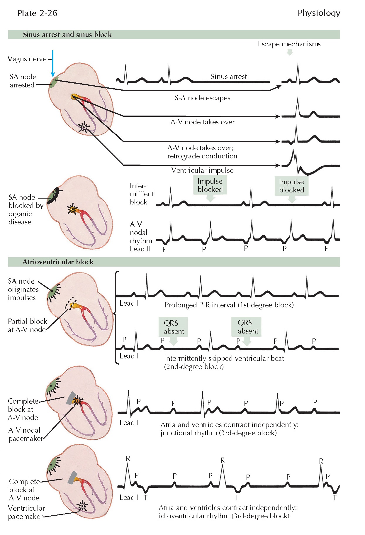
Sinus arrest is usually a functional condition in which the sinus node fails to send impulses to the atria, resulting in a period of cardiac asystole. Eventually, recovery occurs. The first beat after the asystole may be a normal sinus beat (known as a sinus escape beat), or the A-V node may take over for the first beat, originating from a pacemaker in the A-V node, called a nodal escape beat (see Plate 2-26). Here, the P wave may be detectable when there is retrograde atrial conduction. In this case, inverted P waves either precede or follow the QRS complexes in leads II, III, and aVF, or there may be no retrograde conduction and hence no P waves. A ventricular escape beat has all the characteristics of a ventricular ectopic beat. Escape beats of these various types may precede sinus rhythm, sinus bradycardia, sinus tachycardia, nodal rhythm, ventricular tachycardia, or other cardiac rhythms or arrhythmias.
In SA block the
sinus node is blocked organically or chemically (see Plate 2-26). The block may occur intermittently, in which case every other beat may
fail to appear. The sinus node recovers slowly after depolarization, and the
refractory period is such that only every other beat is written. Periodically,
in some cases, more than one beat is skipped. In other cases the SA node is
more severely diseased, and repolarization of the P wave occurs slowly. An A-V
nodal rhythm develops and takes over the role of pacemaker, and the P waves are
inverted in leads II, III, and aVF.
Atrioventricular
block often is classified as a first-, second-, or third-degree A-V block (see Plate 2-26). A first-degree A-V block has a
prolonged P-R interval. A second-degree A-V block is characterized by
the occasional dropping of a QRS complex. A third-degree A-V block shows
a complete dissociation between atrial and ventricular contractions. With a
first-degree A-V block, the P-R interval is long, exceeding 0.2 second for
rates above 60 beats/min. With a second-degree A-V block, an occasional P wave
is not followed by a QRS complex. Normally there is one P wave for every QRS
complex, but with a second-degree block, there may be seven P waves for every
six QRS complexes (or some other ratio of P to QRS). This degree of block would
be designated 7 : 6. A complete A-V block may be of two types: (1) the
pacemaker may be in the A-V nodal tissue, with QRS complexes essentially normal
in appearance and not wide, or (2) the pacemaker may be in His Purkinje system,
and the QRS complexes will be wide and abnormal in shape. In either case, there
will be a complete dissociation between the beating of the atria and the
ventricles. There also will be two different frequencies: one frequency of
about 76 beats/min, which represents the atrial depolarization rate, and the
other 30 beats/ min, which is the ventricular-depolarization rate.
These
arrhythmias are important clinically because a slow rate from any cause greatly
decreases cerebral, coronary, and other organ circulation, which results in
tissue damage and often death. Every medical or surgical effort should be made
to maintain a normal rate. Stopping drugs such as beta blockers and calcium
antagonists or initiating pacemaker therapy may be of great benefit.


