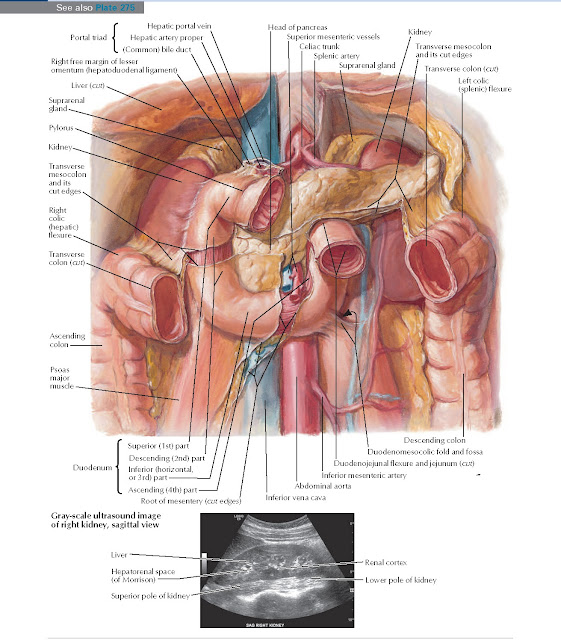
Duodenum in Situ
Anatomy
Suprarenal gland, Kidney, Transverse mesocolon and its cut edges, Transverse colon (cut), Left colic (splenic) flexure, Head
of pancreas, Superior
mesenteric vessels, Celiac
trunk, Splenic artery, Hepatic portal vein, Portal triad, Gray-scale ultrasound image of right kidney, sagittal view, Hepatic artery proper, (Common) bile duct, Right free margin of lesser omentum (hepatoduodenal ligament),
Liver (cut), Suprarenal gland, Pylorus, Kidney, Transverse mesocolon and its cut edges, Right colic (hepatic) flexure, Transverse colon (cut), Ascending colon Psoas major Muscle, Superior (1st) part, Descending (2nd) part, Inferior (horizontal, or 3rd) part, Ascending (4th) part, Root of mesentery (cut edges), Duodenum, Liver, Renal cortex, Lower
pole of kidney, Hepatorenal
space (of Morrison), Superior pole of kidney, Descending colon, Duodenomesocolic fold and fossa, Duodenojejunal flexure and
jejunum (cut), Inferior
mesenteric artery, Abdominal
aorta, Inferior vena cava.


