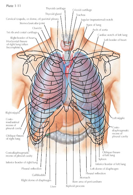
Topography Of The Lungs (Anterior
View)
Because the apex of
each lung reaches as far superiorly as the vertebral end of the first rib, the
lung usually extends about 1 inch above the medial third of the clavicle when
viewed from the front. Thus, the lung projects into the base of the neck.
The anterior border of the right lung descends behind the
sternoclavicular joint and almost reaches the midline at the level of the
sternal angle. It continues inferiorly posterior to the sternum to the level of
the sixth chondrosternal junction. There the inferior border curves laterally
and slightly inferiorly, crossing the sixth rib in the midclavicular line and
the eighth rib in the midaxillary line. It then runs posteriorly and medially
at the level of the spinous process of the tenth thoracic vertebra. These
levels are, of course, variable and apply to the lung in expiration. In
inspiration, the levels for the inferior border are roughly two ribs lower. The
anterior border of the left lung is similar in position to that of the right
lung. However, at the level of the fourth costal cartilage, it deviates
laterally because of the heart, causing a cardiac notch in this border of the
lung. The inferior border of the left lung is similar in position to that of
the right lung except that it extends farther inferiorly because the right lung
is pushed up by the liver below the diaphragm on the right side.
The oblique fissure of the right lung, separating the lower lobe from the
upper and middle lobes, ends at the lower border of the lung near the midclavicular
line. The horizontal fissure separating the middle from the upper lobe begins at
the oblique fissure and runs horizontally forward to the lung’s anterior border,
which it reaches at about the level of the fourth costal cartilage.
The oblique fissure of the left lung is similar in its location to the
corresponding fissure of the right side. The left lung ordinarily has only two
lobes, and there is usually no horizontal fissure in this lung. Extra fissures
may occur in either lung, usually between bronchopulmonary segments and, in the
left lung, between the superior and inferior divisions of the upper lobe,
giving rise to a three-lobed left lung.
The lungs seldom extend as far inferiorly as the parietal pleura, so some
of the diaphragmatic parietal pleura is usually in contact with costal parietal
pleura. This
Area which, of course, varies in size with the phase of respiration is
called the costodiaphragmatic recess of the pleura or the costophrenic
sulcus. A similar but much less extensive area is present where the
anterior border of the lung does not extend to its limits medially especially
in expiration and the costal and mediastinal parietal pleurae are in contact.
This area is called the costomediastinal recess.
The diaphragm separates the liver from the right lung and, depending on
the size of the liver, from the left lung. The left lung is also separated by
the diaphragm from the stomach and the spleen.
The nipple in males usually overlies the fourth intercostal space in
approximately the midclavicular line. In females, its position varies,
depending on the size and functional
state of the breast.


