Lungs Anatomy
The two lungs lie in
the thoracic cavity and are separated by the structures in the mediastinum (Fig. 2.19). Although the lungs of infants are pink, those
of older individuals may have a mottled appearance due to deposits of inhaled
carbon. Living lungs are elastic, enabling their volumes to change during
ventilation, in contrast to embalmed lungs, which are rigid and often bear
the imprints of adjacent structures. Each lung is covered in visceral pleura
and is cone-shaped, with the base or diaphragmatic surface directed downwards
and the apex upwards. The costal surface is smoothly convex, while the mediastinal
surface is irregular and bears the hilum of the organ. Fissures are usually
present, and divide each lung into lobes (usually three lobes on the right and
two on the left). Most of the lung consists of the peripheral part of the
respiratory tract and the associated pulmonary vascular system. Having entered
the lung, the bronchi and pulmonary vessels subdivide extensively (Fig. 2.26).
Although variations occur, each lung
is usually divided into upper and lower lobes by an oblique fissure. On the
right, the upper lobe is further subdivided by the horizontal fissure (Fig.
2.20), which runs from the anterior border of the lung into the oblique fissure
and demarcates the middle lobe. On the left, the horizontal fissure is usually
absent and the middle lobe is represented by the lingula (Fig.
2.21).
Surfaces, borders and relations
The costal surface is convex and
extends upwards into the cervical part of the pleura to form the apex of the
lung, which is closely related to the corresponding subclavian artery and vein.
The inferior surface (base) is markedly concave (Figs 2.22
& 2.23), conforming to the upward convexity of the dome of the
diaphragm. The costal and diaphragmatic surfaces meet at the sharp inferior
border. The anterior border is also sharp and is formed where the costal and
mediastinal surfaces are in continuity. In contrast, the posterior border is
rounded and rather indistinct.
Each lung is attached to the
mediastinum by the lung root, the principal components of which are the pulmonary
vessels and the bronchi. These structures, accompanied by bronchial vessels,
lymphatics and autonomic nerves, enter or leave the lung through the hilum.
Usually, two pulmonary veins emerge from each lung, the inferior vein being the
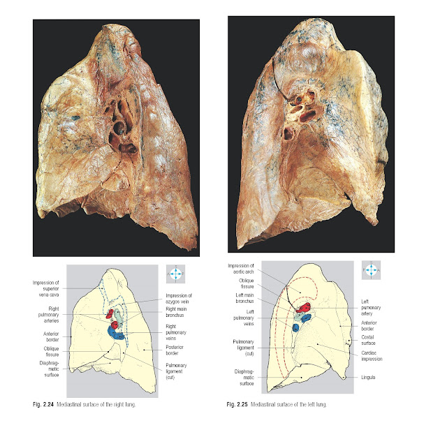
lowest structure in the hilum (Figs 2.24 & 2.25). The bronchi and pulmonary arteries are adjacent as
they pass through the hilum, and on the left, the main bronchus lies
posteroinferior to the pulmonary artery. However, on the right, the main
bronchus frequently divides into two branches, the upper and lower lobe
bronchi, before reaching the lung, and each bronchus is accompanied by a branch
of the pulmonary artery. The hila of both lungs often contain lymph nodes, which
are recognizable by their acquired dark coloration (Fig. 2.27).
The two
lungs have different medial relations. On the right, the anterior part of the
mediastinal surface of the lung is related to the right brachiocephalic vein,
the superior vena cava and the pericardium covering the right atrium of the
heart. Intervening between these structures and the mediastinal pleura is the
right phrenic nerve, which descends in front of the hilum to reach the
diaphragm. The upper part of the hilum is related to the azygos vein (Fig. 2.24),
which arches forwards to terminate in the superior vena cava. The trachea and
accompanying right vagus nerve are related to the right upper lobe.
On the left, the mediastinal surface
of the lung bears distinct impressions produced by the fibrous pericardium and
the heart (Fig. 2.25). The left phrenic nerve is related to the mediastinal
pleura and passes in front of the hilum as it descends across the pericardium.
The aorta creates an obvious groove (Fig. 2.25) where it arches over the lung
root and descends behind the hilum as the descending thoracic aorta.
Surface markings
The apex of each lung rises above the
medial third of the clavicle. From here the anterior border of the lung follows
the reflection of the parietal pleura, passing behind the sternoclavicular and
manubriosternal joints. On the right, the border descends vertically, close to
the midline from the level of the second to the sixth costal cartilages (Fig.
2.2). On the left, the heart displaces the lung and parietal pleura so that the
pericardium is exposed behind the medial ends of the fourth and fifth
intercostal spaces. This location is sometimes used to insert a needle into the
pericardial cavity or heart. On both sides, the inferior border of the lung
crosses the sixth rib in the midclavicular line, the eighth rib in the
midaxillary line and the tenth rib 5 cm from the midline posteriorly. The lower
border of the lung lies at a higher level than the line of pleural reflection;
this part of the pleural cavity not occupied by lung is called the
costodiaphragmatic recess (Fig. 4.105) and may be fluid filled in pleural
effusion.


