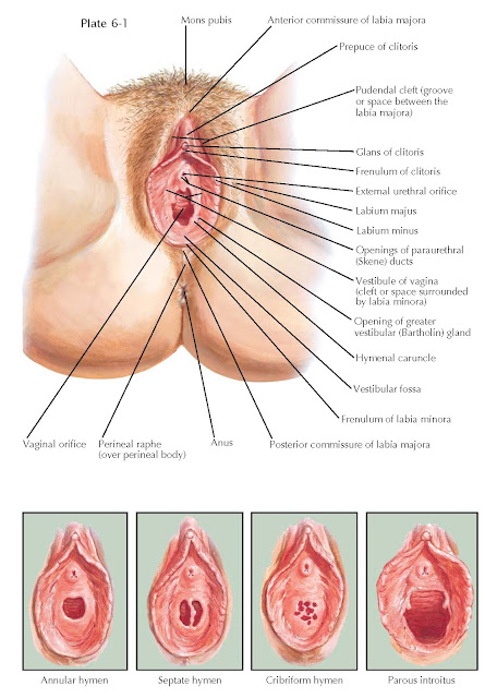
EXTERNAL GENITALIA
The vulva includes those portions of the female genital tract that are externally visible in the perineal region. The mons veneris, overlying the symphysis pubis, is a fatty prominence, covered by terminal sexual (pubic) hair that functions as a dry lubricant during intercourse. From the mons, two longitudinal folds of skin, the labia majora, extend in elliptical fashion to enclose the vulval cleft. They contain an abundance of adipose tissue, sebaceous glands, and sweat glands and are covered by hair on their upper outer surfaces. The anterior commissure marks their point of union at the mons. Posteriorly, a slightly raised connecting ridge, the posterior commissure or fourchette, joins them. Between the fourchette and the vaginal orifice, a shallow, boat-shaped depression, the fossa navicularis, is evident. The labia minora are thin, firm, pigmented, redundant folds of skin, which anteriorly split to enclose the clitoris; laterally, they bound the vestibule and diminish gradually as they extend posteriorly. The skin of the labia minora is devoid of hair follicles, poor in sweat glands, and rich in sebaceous glands. The skin of the labium majus, and to a less extent the labium minus, is subject to most of the same dermatologic pathologies as other areas of skin.
The clitoris, a small, cylindrical, erectile organ situated at the lower
border of the symphysis is composed of two crura, a body, and a glans. The
crura lie deeply, in close apposition to the periosteum of the ischiopubic
rami. They join to form the body of the clitoris, which extends downward
beneath a loose prepuce to be capped by the acorn-shaped glans. Only the glans
of the clitoris is generally visible externally between the two folds formed by
the bifurcation of the labia minora. When the clitoris is abnormally enlarged
as a result of exposure to excess androgens, the clitoral index (the product of
the sagittal and transverse diameters of the glans, in millimeters; normal 35
mm2) is used to grade the degree of enlargement.
The vestibule becomes apparent on separation of the labia. Within it are
found the hymen, the vaginal orifice, the urethral meatus, and the opening of
Skene and Bartholin ducts. The external urethral meatus is situated upon a
slight papilla-like elevation, 2 cm below the clitoris. In the posterolateral
aspect of the urinary orifice, the openings of Skene ducts lie. They run below
and parallel to the urethra for a distance of 1 to 1.5 cm. Bartholin ducts are
visible on each side of the vestibule, in the groove between the hymen and the
labia minora, at about the junction of the middle and posterior thirds of the lateral boundary of the vaginal orifice. Each duct, approximately
1.5 cm in length, passes inward and laterally to the deeply situated vulvovaginal
glands. The Bartholin glands are situated posterior to the 3- and 9-o’clock
locations, which is important clinically when a Bartholin gland abscess is
considered in patients with labial swelling.
The hymen is a thin, vascularized membrane that separates the vagina from
the vestibule. It is covered on both sides by
stratified squamous epithelium. As a rule, it shows great variations in
thickness and in the size and shape of the hymenal openings (e.g., annular,
septate, cribriform, crescentic, fimbriate). After tampon usage, coitus, and
childbirth, the shrunken remnants of the hymen are known as carunculae
hymenales or hymenal caruncles. The presence or absence of an intact hymen is
insufficient to de ermine the presence or absence of past sexual activity.


