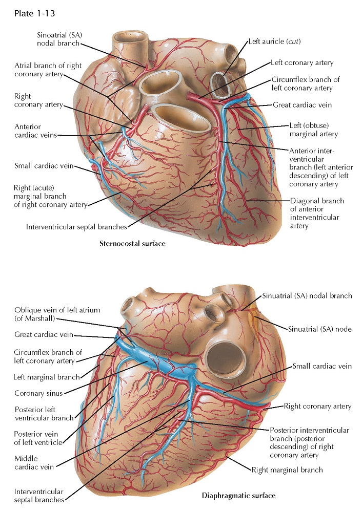
Coronary Arteries and Cardiac Veins
 |
| STERNOCOSTAL AND DIAPHRAGMATIC SURFACES |
The normal heart and the proximal portions of the great vessels receive their blood supply from two coronary arteries. The left coronary artery (LCA) originates from the left sinus of Valsalva near its upper border, at about the level of the free edge of the valve cusp. The LCA usually has a short (0.5-2 cm) common stem that bifurcates or trifurcates. One branch, the anterior inter- ventricular (descending) branch, courses downward in the anterior interventricular groove (largely embedded in fat), rounds the acute margin of the heart just to the right of the apex, and ascends a short distance up the posterior interventricular groove.
The left anterior descending branch
of the LCA gives off branches to the adjacent anterior RV wall (which usually
anastomose with branches from the right coronary artery) and septal branches
(which supply anterior two thirds and apical portions of septum), as well as a
number of branches to the anteroapical portions of the left ventricle,
including the anterior papillary muscle.
One septal branch originating from
the upper third of the anterior interventricular branch is usually larger than
the others and supplies the midseptum, including the bundle of His and bundle
branches of the conduction system. This branch also may supply the anterior
papillary muscle of the right ventricle through the moderator band. The second,
usually smaller circumflex branch of the left coronary artery runs in
the left AV sulcus and gives off branches to the upper lateral left ventricular
wall and the left atrium. The circumflex branch usually terminates at the
obtuse margin of the heart, but it can reach the crux (junction of posterior
inter-ventricular sulcus and posterior AV groove). In this case the circumflex
branch supplies the entire left ventricle and ventricular septum with blood,
with or without the right coronary artery.
In cases where the LCA trifurcates,
the third branch, coming off between the anterior interventricular and the circumflex
branches, is merely an LV branch that originates from the main artery.
The right coronary artery (RCA)
arises from the right anterior sinus of Valsalva of the aorta and runs along
the right AV sulcus, embedded in fat. The RCA rounds the acute margin to reach
the crux in the majority of cases, and it gives off a variable number of
branches to the anterior RV wall. A usually well-developed
and large branch runs along the acute margin of the heart. The posterior
interventricular (descending) branch descends along the posterior
interventricular groove, not quite reaching the apex, and supplies the
posterior third or more of the interventricular septum. The diaphragmatic part
of the right ventricle is largely supplied by small, parallel branches from the
marginal and posterior descending arteries, not from the parent vessel itself. The latter generally crosses the crux,
giving off the posterior interventricular branch and a small branch to the
atrioventricular node. It terminates in a number of branches to the LV wall.
The posterior papillary muscle of
the left ventricle usually has a dual blood supply from both the left and the
right coronary artery.
Of the right atrial branches of
the right coronary artery, one is of great importance. This branch
originates from the RCA shortly after its takeoff and ascends along the
anteromedial wall of the right atrium. It enters the upper part of the atrial
septum, reappears as the superior vena cava branch (nodal artery)
posterior and to the left of the SVC ostium, rounds the ostium, and runs close
to (or through) the sinoatrial node (see Plate 1-13), giving off
branches to the crista terminalis and pectinate muscles.
Variations in the branching pattern
are extremely common in the human heart. In about 67% of cases the RCA crosses
the crux and supplies part of the LV wall and the ventricular septum. In 15% of
cases (as in dogs and many other mammals) the LCA circumflex branch crosses the
crux, giving off the posterior interventricular branch and supplying the entire
left ventricle, the ventricular septum, and part of the RV wall. In about 18%
of cases, both coronary arteries reach the crux. No real posterior
interventricular branch may exist, but the posterior septum is penetrated at
the posterior interventricular groove by many branches from the LCA, RCA, or
both. In about 40% of cases the SVC branch is a continuation of a large anterior
atrial branch of the LCA rather than of the anterior atrial branch of the
RCA.
Also, the first branch of the RCA
may originate independent of the right sinus of Valsalva rather than from the
parent artery. Rarely, the second or even the third RCA branch arises
independently.
Most of the cardiac or coronary
veins enter the coronary sinus. The three largest veins are the great
cardiac vein, middle cardiac vein, and posterior left ventricular vein.
The ostia of these veins may be guarded by fairly well-developed unicuspid or
bicuspid valves. The oblique vein of the left atrium (of Marshall)
enters the sinus near the orifice of the great cardiac vein, and its ostium
never has a valve. The small cardiac vein may enter the right atrium independently, and the anterior
cardiac veins always do.
Small venous systems in the atrial
septum (and probably in ventricular walls and septum) enter the cardiac
chambers directly, called the thebesian veins. The existence of so-called arterioluminal
and arteriosinusoidal vessels is debatable and the evidence inconclusive.


