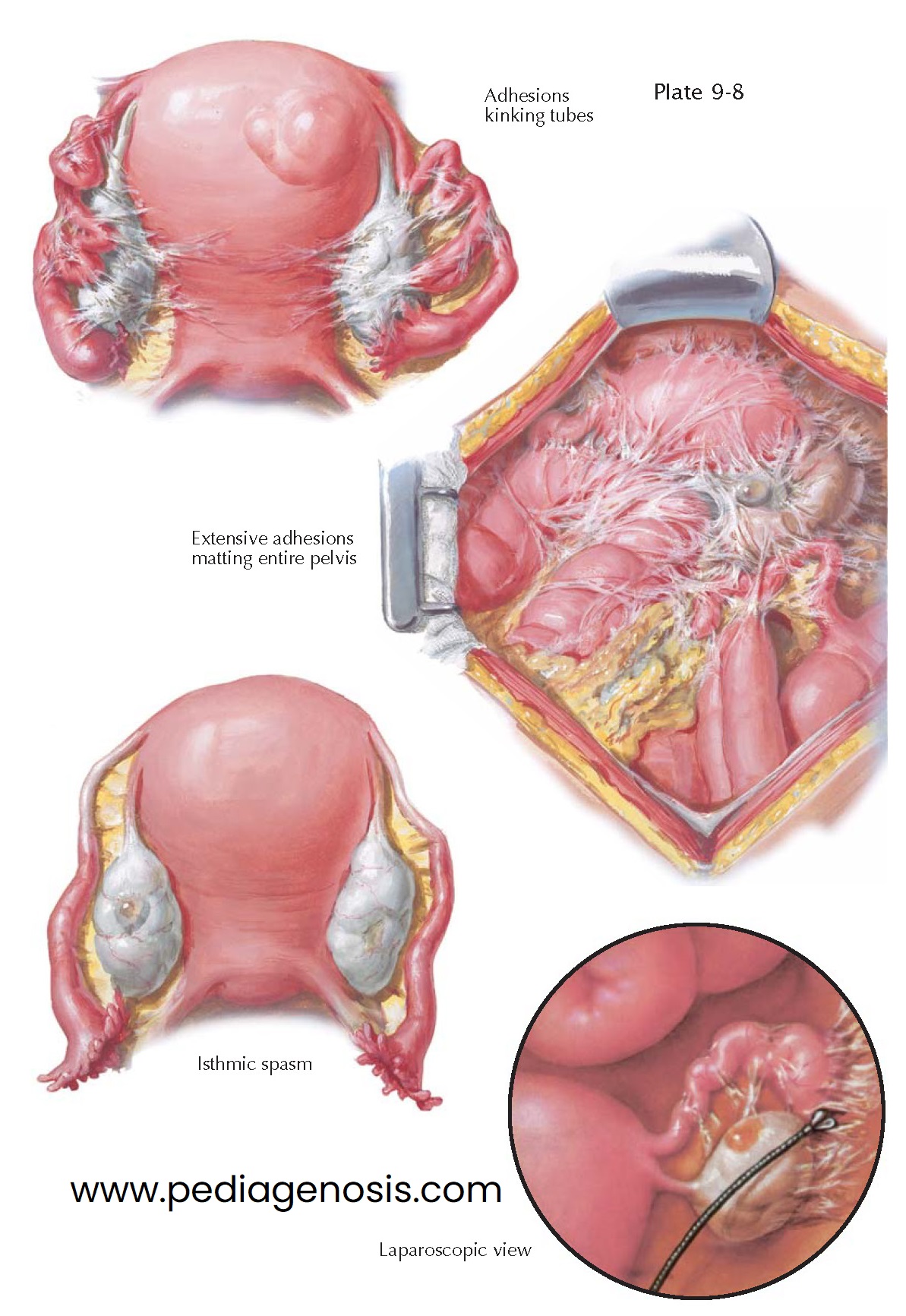
CHRONIC SALPINGITIS, ADHESIONS
In chronic salpingitis,
the uterine tubal ostium is often obliterated, and the tube cannot be visualized
by hysterosalpingography. This must be differentiated from spasms of the isthmic
portion of the tube, which are frequently encountered and offer resistance to uterotubal
insufflation, which can be overcome using moderate pressure or at the time of laparoscopy
under general anesthesia.
Peritoneal adhesions connecting the tube with the ovary and the posterior leaf of the broad ligament may kink the tube and thus cause sterility. They are recognizable on hysterosalpingography by a characteristic spill pattern. Frequently, these pelvioperitonitic adhesions involve all pelvic organs, including the omentum and low intestinal loops. The adhesions are richly vascular at first, but gradually they become poorly vascularized, frail, and spiderweb-like. Only in rare cases may they disappear entirely.
The most conspicuous symptom of chronic salpingitis is pain, which may be
continuous or elicited by stress, defecation, and intercourse and may be aggravated
by the hyperemia and swelling of the premenstrual phase. Dysmenorrhea may also occur,
but more frequently the menstrual flow relieves the pain, and the patient feels better
during and after menstruation. Dys- pareunia is frequent, and intermenstrual pain
(mittel-schmerz) is occasionally observed.
Women with chronic adnexitis are usually sterile. If the tubes become finally
patent again, ectopic pregnancy often occurs owing to impaired peristalsis,
stenosis, loss of normal ciliary function, and kinks in the tubes. Assisted
reproduction techniques that bypass the damaged tube can result in conception, but
these patients tend to continue to have higher than normal pregnancy loss rates.
The differential diagnosis between an inflammatory adnexal tumor and a parametritic
infiltrate is not always easy. The adnexal tumor has convex outlines, whereas
the surfaces of the parametritic infiltrate are concave. In combined diseases of
the adnexa and the parametrium, the palpable tumor is convex above and concave
below, and its broad base is attached to the lateral pelvic wall. An adnexal tumor
can be separated from the pelvic wall by the examining finger, whereas parametritic
infiltrates are frequently in close contact with the wall structures. Ultrasonography
may only demonstrate cystic loculations making it difficult to differentiate
between ovarian cysts, consolidated adnexal damage, and entrapped omentum and bowel.
Ectopic pregnancy may develop in a chronically dis- eased tube. The menstrual
history in such instances is often not characteristic. Tenderness of the palpated
tumor, increased erythrocyte sedimentation rate, and moderate leukocytosis are common
for both ectopic pregnancy and adnexal tumor. A positive pregnancy test
indicates pregnancy, but a negative test does not always disprove it. Rapid increase
in the size of the tumor, in spite of normal temperature, and development of severe
unilateral colicky pain or of sudden shock speak for ectopic pregnancy. Ultrasonography
may demonstrate decidual changes in the uterine cavity but no gestational sac. A
gestational sac may or may not be seen in the adnexa.
Very difficult is the differentiation between a tubal carcinoma and an inflammatory
adnexal tumor. With no history of inflammatory disease, the presence of tubal malignancy
is probable. A blood-tinged or ambercolored serous discharge is suspicious for tubal
carcinoma but may easily be mistaken for a hydrops tubae profluens. A definite diagnosis
is usually possible only after microscopic examination following surgery.
History, bilaterality, and the demonstration of gonococci facilitate the differentiation between salpingitis in the course of a gonorrheal or puerperal infection and appendicitis. A tumor on the right side connected with the uterus does not disprove a primary appendicitis with secondary infection of the right tube. In the differential diagnosis between chronic appendicitis and chronic salpingitis, the exact localization of the most tender spot of the abdominal wall and visualization of the appendix on computed tomography or magnetic resonance imaging may prove to be helpful. So etimes one must resort to laparoscopy or laparotomy.


