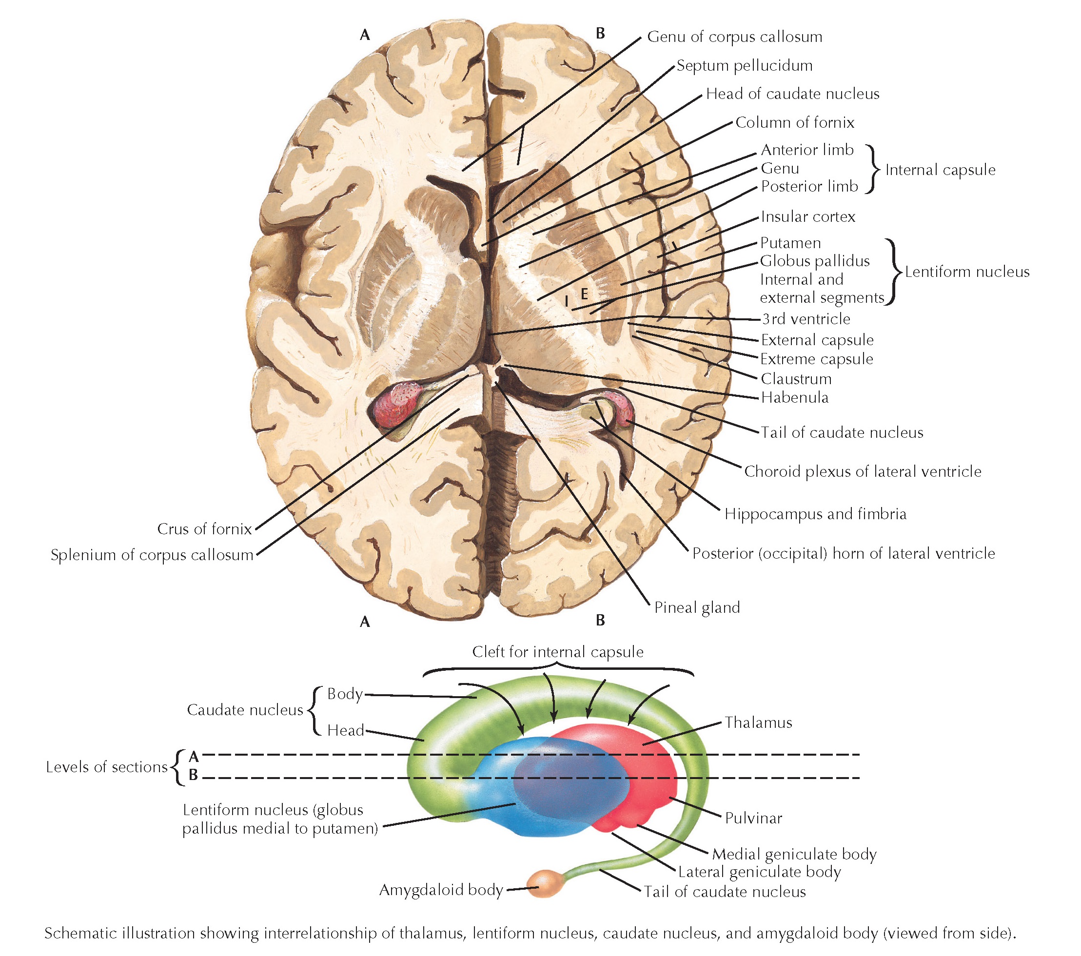
Two levels of horizontal sections through the forebrain reveal the major anatomical features and the relationships among the basal ganglia, the internal capsule, and the thalamus (schematically shown in the lower illustration). The caudate nucleus is a C-shaped structure that sweeps from the frontal lobe into the temporal lobe; a horizontal section passes through this nucleus in two distinct places (head and tail). The anterior limb, genu, and posterior limb of the internal capsule contain major connections into and out of the cerebral cortex. The head and body of the caudate are medial to the anterior limb, whereas the thalamus is medial to the posterior limb. These relationships are important for understanding imaging studies and for understanding the involvement of specific functional systems in vascular lesions or strokes. The internal and external segments of the globus pallidus are located medial to the putamen. The external capsule, claustrum, extreme capsule, and insular cortex, from medial to lateral, are located lateral to the putamen. The fornix, also a C-shaped bundle, is sectioned in two sites, the crus and the column.
The basal ganglia (caudate nucleus, putamen, and globus pallidus) form characteristic anatomical relationships with the internal capsule. The head and body of the caudate nucleus are found medial to the anterior limb; the thalamus is found medial to the posterior limb; and the globus pallidus and putamen are found lateral to the anterior and posterior limbs. Basal ganglia disorders are characterized by movement disorders, although emotional and cognitive symptoms also are seen. Some movement disorders involve actual degeneration of basal ganglia and related structures; these disorders include Huntington’s chorea and degeneration of the head of the caudate nucleus as well as Parkinson’s disease and degeneration of the dopaminergic pars compacta of substantia nigra. Other movement disorders involve altered inhibitory and excitatory activity of specific portions of basal ganglia circuitry; reordering this circuitry may require pharmacologic treatment, therapeutic ablation procedures, or deep brain stimulation.


