 |
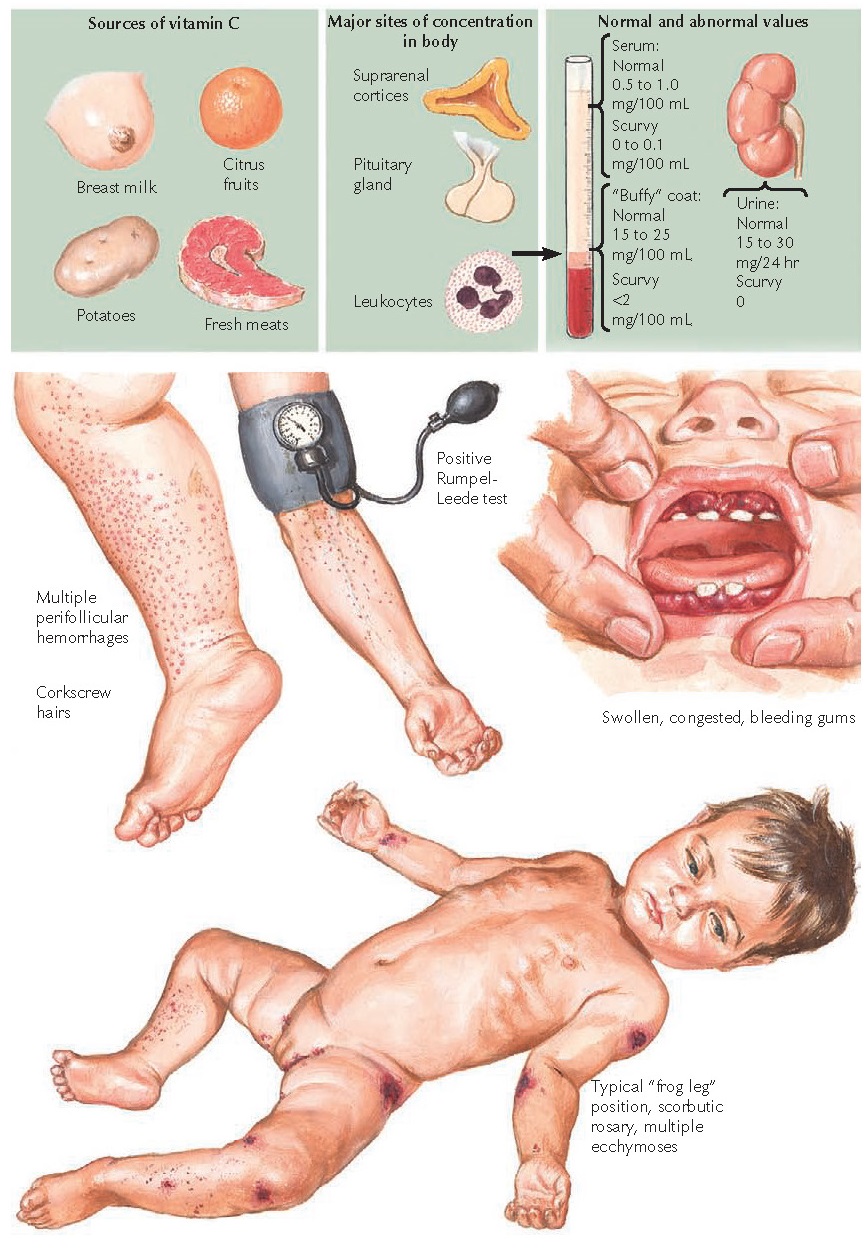
| DIETARY SOURCES OF VITAMIN C AND CLASSIC CUTANEOUS MANIFESTATIONS OF SCURVY |
Scurvy is a well-known nutritional disease that results from a lack of the water-soluble vitamin, ascorbic acid (vitamin C). Scurvy has a well-documented history. It was first recognized in the fourteenth century in sailors who spent long amounts of time at sea. The symptoms were recognized as being related to a lack of fresh foods, especially citrus products. In 1753, James Lind, a British surgeon aboard the HMS Salisbury, performed the first documented clinical trial proving that scurvy was caused by a lack of citrus fruit in the diet of sailors. After Lind’s discovery, citrus fruits were included in ships’ provisions, and the incidence of scurvy in sailors plummeted. It was not until 1928 that ascorbic acid was isolated by the Hungarian chemist, Albert von Szent-Grörgyi, who was eventually awarded the Nobel Prize for this discovery. Scurvy is still present in some areas of the world due to inadequate dietary intake of vitamin C. Scurvy is uncommon in North America but can be seen in individuals with abnormal diets.
Clinical Findings: Scurvy is a disease that can affect a wide range
of organ systems. The skin and mucous membranes are always involved and may
display the initial symptoms of the disease. Recognition of these symptoms is
critical in diagnosing the disease and preventing long-term illness. Scurvy is
a rare disease in regions of the world with access to proper dietary intake of
vitamin C. In North America and Europe, most cases are the result of abnormal
dieting, psychiatric illness, or alcoholism. Prompt recognition of the
cutaneous manifestations can lead to treatment and cure of the disease. Scurvy
has an insidious onset with nonspecific constitutional symptoms such as
generalized weakness, malaise, muscle and joint aches, and easy fatigability
with shortness of breath. These symptoms may be related to the macrocytic
anemia that is frequently seen in patients with scurvy and is believed to be
caused by a coexisting folic acid deficiency.
The
first clinical findings are often in the mucous membranes and the skin.
There are a multitude of cutaneous manifestations. Early in the course of
disease, skin becomes dry and rough, in association with a dulling of the skin
tone. Small, hyperkeratotic papules may be noticed and resemble those of
keratosis pilaris. More specific and sensitive skin findings then develop, including
perifollicular hemorrhage and “corkscrew hairs.” The corkscrew hairs are most
noticeable on the extremities. Swan-neck deformity of the extremity hair may
also occur due to abnormal bending of the hair; this is less common than
corkscrew hair. The nail bed shows splinter hemorrhages. All cutaneous findings
appear to be more common on the lower extremities. This is believed to be a
result of increased hydrostatic pressure in the lower extremities while one is
upright, which leads to increased pressure on the small venules in the
follicular locations, resulting in the perifollicular hemorrhages. These
findings are also observed in areas of pressure directly on the skin, such as
around the waist line. The Rumpel-Leede sign is positive: When a blood pressure
cuff is inflated for 1 minute to a value that is greater than the diastolic
pressure but less than the systolic pressure, numerous petechial hemorrhages
occur distal to and underneath the blood pressure cuff. This test is a sign of
capillary fragility induced by increased hydrostatic pressure.
The
mucous membranes may show the first sign of the disease. The main
finding is edematous, bleeding gums. As the disease progresses, the gums become
friable and peel away from the teeth. The teeth may develop dental calculi
at the base. This may result in loose teeth and pain. Teeth eventually become
disrupted from their attachments and fall out.
Compared
with scurvy in adults, congenital scurvy and scurvy during early childhood have
unique manifestations related to bony development. Vitamin C is critically
important for the development of collagen and cartilage, and abnormalities at a
young age result in a variety of bony deformities. Scorbutic rosary is
a term given to the prominence of the costochondral junctions. Infants with
scurvy develop “frog legs” due to subperiosteal hemorrhage. This form of
hemorrhage is painful, and the infant naturally relaxes the lower limbs in this
pattern to relieve the pain. Healing of the subperiosteal hemorrhage often
involves abnormal calcification of the region and the formation of a more
club-shaped bone.
This
can lead to difficulty with movement. Radiographs of the long bones reveal the
classic white line of Frankel, which represents the abnormal calcification of
the cartilage within the epiphysial-diaphysial juncture. The periosteum appears
ballooned out due to the presence of subperiosteal hemorrhage. Over time, the
hemorrhagic areas become partially or completed calcified.
Infants
with scurvy may also develop severe ecchymosis around the eye and in the
retrobulbar space, which, when severe, can result in proptosis. Child abuse may
be considered in the differential diagnosis.
Breast
milk contains adequate amounts of vitamin C, so infantile scurvy is more likely
to occur in children who are not breast fed and are given a diet devoid of
vitamin C.
Pathogenesis: Vitamin C is an essential vitamin that is acquired
through dietary intake. Humans lack the enzyme L-gluconolactone
oxidase, which is required for the synthesis of L-ascorbic acid from
its precursor, glucose. Dietary sources of vitamin C include fruits,
vegetables, and fresh meats. Citrus fruits are the main source of dietary
vitamin C. All human tissues contain vitamin C, with the adrenal glands and pituitary
glands having the highest concentrations. Leukocytes contain appreciable
amounts of vitamin C, and the buffy coat level is helpful in diagnosis. The
clinical manifestations of scurvy do not appear until the buffy coat
concentration has fallen to less than 4 mg/100 mL or the serum level to less
than 20 µmol/L. The normal buffy coat concentration is in the range of 15 to 25
mg/100 mL, and that of the serum is 40 to 120 µmol/L. The kidney has an
extraordinary ability to adjust its vitamin C reabsorption and secretion based
on serum levels. In scurvy, the kidney salvages all available vitamin C, and
the urine concentration is 0 mg/24 hours.
Vitamin
C is required as a cofactor for various enzyme functions. Vitamin C supplies
electrons to enzymatic reactions. If these are absent, the enzymes are unable
to properly produce their intended end product, and the manifestations of
scurvy begin to develop. One of the most important functions of vitamin C is to
serve as a cofactor, along with ferrous iron (Fe++), for the enzymes prolylhydroxylase
and lysylhydroxylase. These enzymes are responsible, respectively, for
hydroxylation of the proline and lysine amino acid residues in collagen. If the
proper ratio of proline and lysine hydroxylation is not present, the collagen
molecule is unable to form a proper triple helix, and its function is
compromised. Defective collagen production is the main deficiency responsible
for the cutaneous signs of scurvy, because collagen is the major structural
protein in blood vessel walls and in the dermis. Vitamin C is also responsible
for electron donation in other enzymatic reactions, including those that
synthesize tyrosine, dopamine, and carnitine.
Histology: Histology is not required for the diagnosis. Biopsy of
a petechial lesion shows perifollicular red blood cell extravasation and a
minimal lymphocytic inflammatory infiltrate. If the specimen includes the area
around a hair follicle, close inspection will reveal a coiled or corkscrew
appearance to the hair follicle. It should be remembered that patients with
scurvy have impaired wound healing: After biopsy without proper therapy, the
freshly incised skin may take weeks to months to heal, and large ecchymoses
typically develop around the biopsy site.
 |
| BONY AND SKIN ABNORMALITIES OF SCURVY |
Treatment: Therapy requires the replacement of vitamin C at a
dosage of 300 to 500 mg daily until the symptoms resolve. Then start the
recommended daily allowance. Patients show rapid improvement. The root
cause must be determined, and if the patient does not respond to therapy, serum
levels should be rechecked. If they are still low, noncompliance with therapy
should be considered. Often, patients with scurvy have an underlying
alcoholism, eating disorder, or psychiatric illness that, if not properly
addressed, will continue to occur. Patients should see a nutritionist, who can
best educate them on the need for a balanced diet and which foods are high in
vitamin C. Alcoholics need to be referred to experts who are adept at treating
this common problem. Supplementation with the daily recommended amounts can be
continued for life, because any excess vitamin C is not stored in the body but
excreted by the kidneys. Supplementation ensures the avoidance of further episodes of scurvy.


