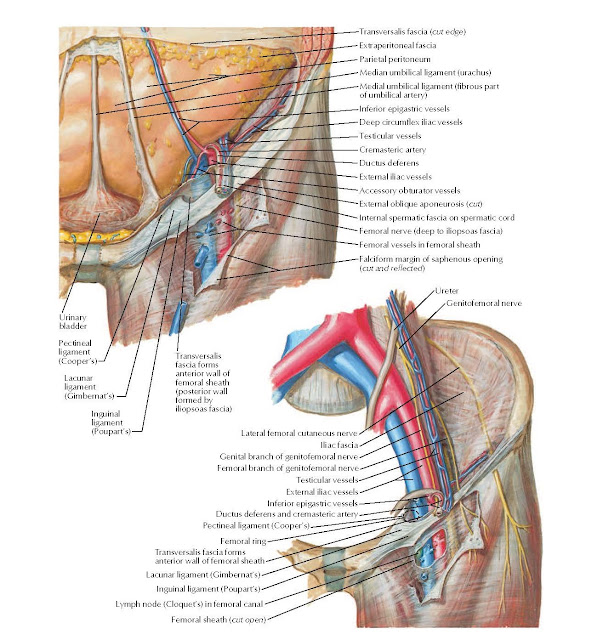
Femoral Sheath and Inguinal Canal Anatomy
Transversalis fascia (cut edge), Extraperitoneal fascia,
Parietal peritoneum, Median
umbilical ligament (urachus), Medial
umbilical ligament (fibrous part of
umbilical artery), Inferior
epigastric vessels, Falciform
margin of saphenous opening (cut
and reflected), Deep
circumflex iliac vessels, Testicular
vessels, Cremasteric artery, Ductus deferens, External iliac vessels, Accessory obturator vessels, External oblique aponeurosis (cut), Internal spermatic fascia on
spermatic cord, Femoral nerve
(deep to iliopsoas fascia), Femoral
vessels in femoral sheath, Transversalis fascia forms anterior wall of femoral
sheath (posterior wall formed by iliopsoas fascia).
Urinary bladder, Ureter, Genitofemoral nerve, Lateral
femoral cutaneous nerve Iliac fascia, Genital branch of genitofemoral
nerve, Femoral branch of
genitofemoral nerve, Testicular
vessels, External iliac
vessels, Inferior epigastric
vessels, Ductus deferens and
cremasteric artery, Femoral
ring, Transversalis fascia
forms anterior wall of femoral sheath, Lymph node (Cloquet’s) in femoral
canal, Femoral sheath (cut
open) Inguinal ligament (Poupart’s), Pectineal ligament (Cooper’s), Lacunar
ligament (Gimbernat’s).


