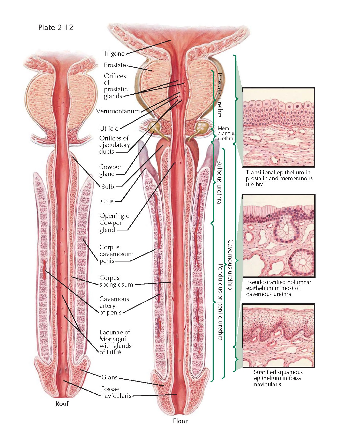
Urethra and Penis
The figure shows the entire length of the male
urethra as it traverses the prostate, pelvic diaphragm, and the penis. The
natural curvature (see Plate 2-1) of the penis has been removed. The fascial
relationships among tissues in the penis are not emphasized here but can be
found in Plates 2-2 through 2-4. The pendulous, or penile urethra, and bulbous
urethra extend through the center of the spongy tissue called the corpus
spongiosum that joins with the paired corpora cavernosa to form the penile
shaft. Each of the three spongy bodies is enclosed in a fibrous capsule, the
tunica albuginea (see lower portion of Plate 2-3). The cavernosal and
spongiosal bodies have a separate blood supply and there are normally no
vascular anastomoses between them. Vascular shunts between these bodies may
occur with trauma, however. The spongy tissue of the corpora cavernosa and
spongiosum is composed of large venous sinuses that become widely dilated and
engorged with blood during penile erection.
The
urethral epithelium varies in different anatomic segments of the urethra. From the bladder neck to
the triangular ligament (pars prostatica), the funnel-shaped coning of the
trigone as it progresses distally into the prostatic urethra, the epithelium is
transitional in character. In the membranous urethra (pars membranacea) that
traverses the urogenital diaphragm, the epithelium assumes a stratified columnar
form. The epithelium of the penile urethra (pars cavernosa) is composed of pseudostratified
and columnar cells. Distally, in the fossa navicularis, the epithelial cells
are stratified squamous in nature. The urethral mucosa is surrounded by the
lamina propria that consists of areolar tissue with venous sinuses and bundles
of smooth, unstriated muscle.
The floor
of the prostatic urethra contains numerous orifices that represent the terminal
ducts of the prostatic acini. Also on the prostatic urethral floor is an obvious
elevation called the verumontanum, colliculus seminalis, or prostatic utricle.
This mound of tissue contains a small pocket or utricle that represents the
fused ends of each of the müllerian ducts (see Plate 1-2). In fact, the utricle
is considered the male remnant of the female uterus. Just distal and lateral to
the utricle are the slit-like orifices of the paired ejaculatory ducts, the obstruction of
which is a well-defined cause of male infertility.
Within
the penile periurethral tissue are many small, branched, tubular glands, the
epithelia of which contain modified columnar, mucus-secreting cells. These glands
of Littré are more numerous in the roof than the floor of the penile urethra.
Also found in the roof of the penile urethra are many small recesses called the
lacunae of Morgagni, into which the glands of Littré empty. These lacunae and
glands may become chronically
infected following urethritis, resulting in recurring symptoms and urethral
discharge.
The
pea-sized bulbourethral glands of Cowper lie laterally and posteriorly to the
membranous urethra between the fasciae and the urethral sphincter within the
urogenital diaphragm (see Plate 2-5). The ducts of these glands, about an inch
long, pass obliquely forward and open on the floor of the bulbous urethra. The
secretions from these glands form part of the seminal fluid during
ejaculation.
Give us your opinion


